How Muscles Rebuild ATP: Phosphocreatine, Glycolysis, and Lactate
How can muscles keep moving with so little stored ATP?
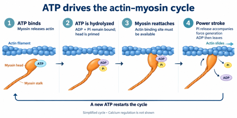
The ATP already stored in a muscle cannot sustain strong contractions for long. As ATP is used, it must also be resynthesized, turning ADP and other inputs back into ATP.
Here, the size of the ATP pool and its turnover rate are different things. The pool is the amount present at one moment. Turnover is the rate at which ATP is used and regenerated over time. Repeated cycling allows the total amount processed to be much larger than the amount present at any single instant.
Estimates of the large mass of ATP used by the body each day illustrate this distinction. They do not mean that the entire amount is stored in the body at once.
When exercise begins, ATP use accelerates, and the pathways that regenerate it increase their activity. This can be understood as metabolic flux, the amount of material passing through a pathway in a given time. The important question becomes: Can ATP be supplied at the rate required? [4]
Phosphocreatine rapidly transfers a phosphate to ADP
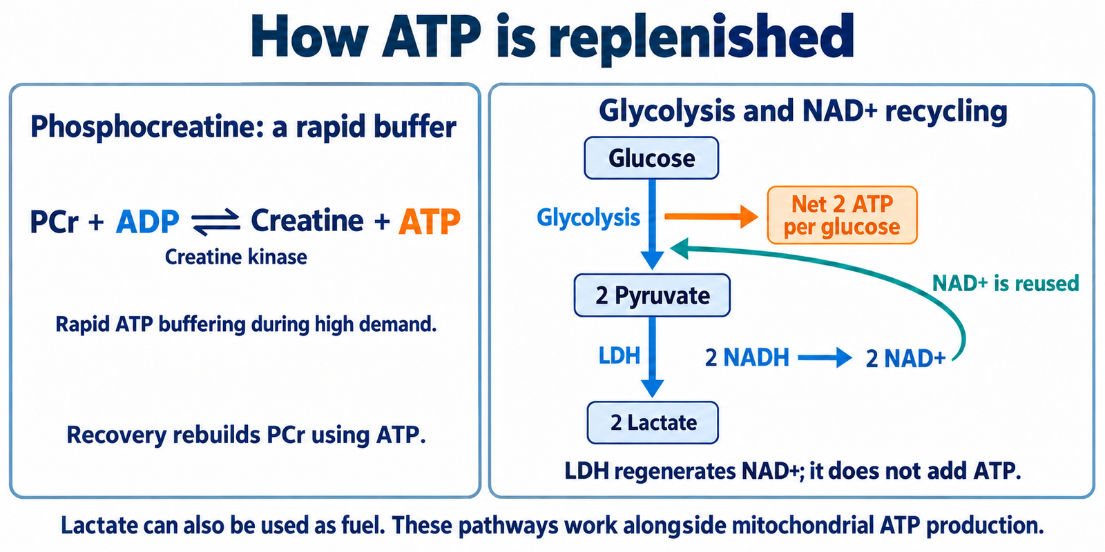
Phosphocreatine, or PCr, consists of a phosphate group attached to the small organic molecule creatine. When muscle ATP demand rises suddenly, PCr can rapidly transfer that phosphate to ADP:
ADP + PCr ⇄ ATP + creatine
The enzyme creatine kinase, CK, accelerates this reaction. This is a simplified equation that omits hydrogen ions and other details.
PCr does not directly power myosin. It regenerates ATP from ADP, and that ATP is then used by myosin and calcium pumps. The pathway acts as a fast buffer, helping stabilize ATP supply when demand changes abruptly.
Its strength is speed; its limitation is the finite size of the PCr store. A rapid reaction cannot provide an unlimited supply.
During recovery, ATP produced by other metabolic pathways drives the reaction in the reverse direction to replenish PCr. PCr replenishes ATP during demand, and ATP production subsequently replenishes PCr. This system is therefore linked to the other energy pathways rather than operating independently. [2, 4]
Glycolysis produces ATP while processing sugar
Glycolysis changes the carbon skeleton of glucose through a series of reactions, producing two molecules of pyruvate, a three-carbon metabolic intermediate. It occurs in the cytosol, the fluid compartment outside the cell’s membrane-bound organelles.
Muscle can use glucose taken up from the blood or break down its own glycogen, a storage molecule made of linked glucose units, to supply material for glycolysis.
For one glucose molecule arriving from the blood, the overall sequence is:
- Two ATP are invested. Phosphate groups are added, preparing the sugar for later reactions.
- The six-carbon framework is split into two three-carbon frameworks.
- Four ATP are produced as intermediates are converted. Subtracting the two initially invested gives a direct net gain of two ATP.
- Two pyruvate and two NADH are formed. NADH is a cofactor that has accepted electrons during the pathway.
ATP is made here by substrate-level phosphorylation. A substrate is a molecule on which an enzyme acts; phosphorylation means adding a phosphate group. In glycolysis, phosphate is transferred directly from a metabolic intermediate to ADP to make ATP.
The direct ATP yield per glucose is small, but increasing glycolytic flux can increase production per unit time. Yield per molecule and production rate per second are different measures. Starting from glycogen can change the initial ATP cost, so the figure of two net ATP must be tied to its starting material: glucose. [4, 6]
Why is lactate made, and where does it go?
Lactate formation is a separate reaction after glycolysis
NAD⁺ is a cofactor that can accept electrons; NADH is its electron-carrying form. Oxidation means loss of electrons, and reduction means gain of electrons.
The final step of glycolysis is the reaction in which pyruvate kinase converts phosphoenolpyruvate, PEP, into pyruvate while producing ATP. Lactate is not an intermediate within that sequence. It is formed from pyruvate in a separate reaction catalyzed by lactate dehydrogenase, LDH. [6, 12]
Pyruvate receives electrons from NADH and is reduced to lactate; NADH is converted back to NAD⁺:
Pyruvate + NADH + H⁺ ⇄ lactate + NAD⁺
If both pyruvate molecules produced from one glucose become lactate:
2 pyruvate + 2 NADH + 2 H⁺ ⇄ 2 lactate + 2 NAD⁺
Glycolysis needs the regenerated NAD⁺
At the glyceraldehyde-3-phosphate dehydrogenase, GAPDH, step of glycolysis, glyceraldehyde-3-phosphate, GAP, is converted to 1,3-bisphosphoglycerate, 1,3-BPG. NAD⁺ accepts electrons and becomes NADH. For that reaction to continue, NAD⁺ must become available again.
The NAD⁺ regenerated by LDH can be reused at this step. Lactate itself does not loop back into the middle of glycolysis to keep it running. Insufficient NAD⁺ regeneration can limit glycolytic flux. [6]
The LDH reaction does not directly produce additional ATP. It regenerates NAD⁺, helping sustain the glycolytic reactions that do produce ATP.
Lactate can fuel muscle and heart
Lactate is not a useless waste product. It can serve as fuel for more oxidative fibers within the same muscle, other muscles, and the heart. The bloodstream carries it between tissues. [7, 11]
To use it as fuel, the reversible LDH reaction converts lactate back into pyruvate. Pyruvate can then become acetyl-CoA and enter the TCA cycle. Electron carriers generated during this metabolism support the electron transport chain and oxidative phosphorylation, allowing ATP production. [7–9]
Lactate → pyruvate → acetyl-CoA → TCA cycle and oxidative phosphorylation → ATP production
A substance produced in one location can therefore supply ATP production elsewhere. Oxidation is a major route of lactate disposal during exercise. Its contribution relative to other routes varies with exercise intensity and metabolic state. [11]
Some lactate is turned back into glucose in the liver
Some lactate travels through the blood to the liver. There it is converted to pyruvate and used in gluconeogenesis, the synthesis of glucose from precursors such as lactate. [12]
The resulting glucose can return to the blood and be used again by muscle. This sequence, muscle lactate → liver glucose production → glucose supplied to muscle, is called the Cori cycle. [12]
The liver must invest energy, including ATP and GTP, to rebuild glucose. This is not a cycle that generates energy for free. Nor does all lactate travel to the liver: oxidation by muscle and heart occurs alongside glucose production in the liver. [11, 12]
Keep these two roles separate
- When lactate is formed: conversion of NADH back to NAD⁺ helps glycolysis continue.
- When lactate is used: it can provide fuel for oxidative metabolism or material for glucose synthesis.
Lactate is produced and used even when oxygen is available. The fact that glycolysis does not directly consume oxygen does not mean lactate forms only in the complete absence of oxygen. Its concentration reflects production, use, and transport together. [7, 11]
References
[2] OpenStax: Muscle Fiber Contraction and Relaxation.
[4] Hargreaves and Spriet: Skeletal Muscle Energy Metabolism During Exercise (2020); Kazak and Cohen: Creatine Metabolism (2020).
[6] OpenStax: Glycolysis.
[7] Brooks: Cell–Cell and Intracellular Lactate Shuttles (2009).
[8] OpenStax: Oxidation of Pyruvate and the Citric Acid Cycle.
[9] OpenStax: Oxidative Phosphorylation.
[11] Brooks: Lactate—Link Between Glycolytic and Oxidative Metabolism (2007).
[12] NCBI Bookshelf: Biochemistry, Gluconeogenesis.
Korean original reviewed for medical accuracy by S. Chang, KMD.
This article is for educational purposes and is not individual medical advice.
Written by: MedMaru Editorial Team
Reviewed for medical accuracy by:
S. Chang, KMD