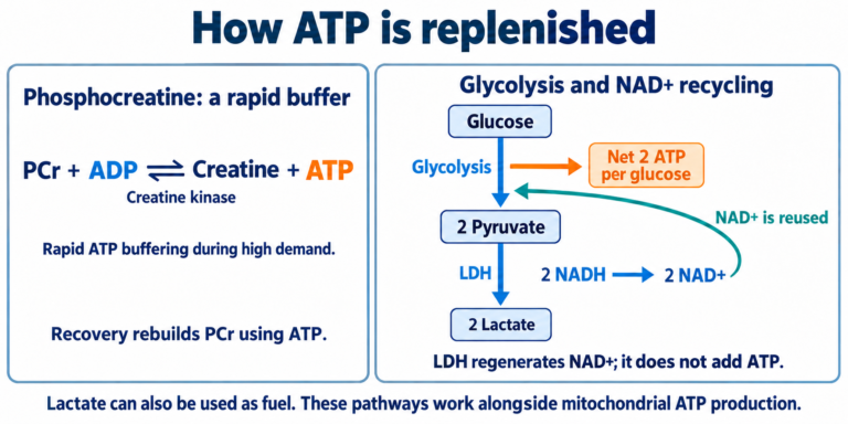
How ATP Powers Muscle Contraction and Relaxation
ATP is often called the cell’s energy currency. To understand what that means, it helps to follow the molecule itself: what it is made of, how it changes during a reaction, and how those changes make a muscle move.
ATP is a molecule with a specific structure
ATP stands for adenosine triphosphate. Its name describes its structure:
- Adenine: a nitrogen-containing, ring-shaped base.
- Ribose: a five-carbon sugar.
- Three phosphate groups: three linked chemical units containing phosphorus and oxygen.
Adenine and ribose together form adenosine. Adding three phosphate groups gives adenosine triphosphate. The phosphates are named alpha (α), beta (β), and gamma (γ), starting with the one closest to ribose. Gamma is the terminal phosphate.
ADP is adenosine diphosphate. It has the same basic framework as ATP but two phosphate groups rather than three. Thinking of ATP and ADP as related molecules that differ in phosphate number makes their cycling easier to understand.
Using ATP does not mean pouring a substance called “energy” out of a container. It means coupling a reaction that changes ATP into other molecules to work that the cell needs to perform. [1]
ATP hydrolysis: why the whole reaction matters
Hydrolysis is a reaction in which water participates in the cleavage of a chemical bond. The ATP reaction discussed here can be written in simplified form:
ATP + water → ADP + Pi
Pi means inorganic phosphate: phosphate that is not attached to another organic molecule. This simplified equation leaves out details of charge and hydrogen ions.
The phrase “breaking ATP’s bond releases energy” can be misleading. Breaking a bond itself requires energy. As ATP and water become ADP and Pi, existing bonds are broken, new bonds form, and interactions with surrounding water change. Whether a reaction is energetically favorable depends on this complete change, not on bond breaking alone.
The quantity that describes this is the change in Gibbs free energy, ΔG. At constant temperature and pressure, it indicates which direction of a reaction is thermodynamically favorable. Under typical cellular conditions, ATP hydrolysis has a negative ΔG, allowing it to help drive other processes.
Under standard biochemical conditions, the value is approximately −30.5 kJ/mol. Inside a cell, however, it varies with ATP, ADP, and Pi concentrations and other conditions. A value around −50 kJ/mol illustrates a possible cellular value; it is not a fixed amount released by ATP under every circumstance.
A favorable reaction is not necessarily a fast reaction. Cells use enzymes, biological catalysts that accelerate reactions, to control ATP use. [1]
How does a chemical reaction become movement?
Muscle generates force by harnessing the free-energy change associated with ATP hydrolysis. The protein that connects this chemistry to movement is myosin.
Coupling means linking an energy-releasing process to a process that requires energy so that the latter can proceed. Here, coupling describes two processes working together, not merely two molecules sticking to one another.
A bicycle offers a useful analogy: force applied to the pedals is transmitted through the chain to the wheels. In a muscle, changes in myosin’s shape provide the connection instead of a chain.
As ATP binds to myosin, is hydrolyzed, and releases its reaction products, myosin changes both its shape and its affinity for actin. These changes allow it to release actin, prepare for another cycle, reattach, and generate force. ATP chemistry and myosin’s mechanical operation are linked. [1, 3]
Contraction: one cycle of the molecular motor
The following explanation concerns skeletal muscle, the muscle that moves the limbs. Two proteins form the main interacting filaments inside its cells:
- Actin is the main protein in the thin filament, which myosin binds to and pulls against.
- Myosin is a molecular motor whose heads bind actin and use ATP to generate force.
The connection through which myosin transmits force to actin is called a cross-bridge. When a muscle shortens, the actin and myosin filaments do not themselves become shorter. They slide past one another, changing how much they overlap.
Because the cross-bridge cycle repeats, we can begin with myosin attached to actin:
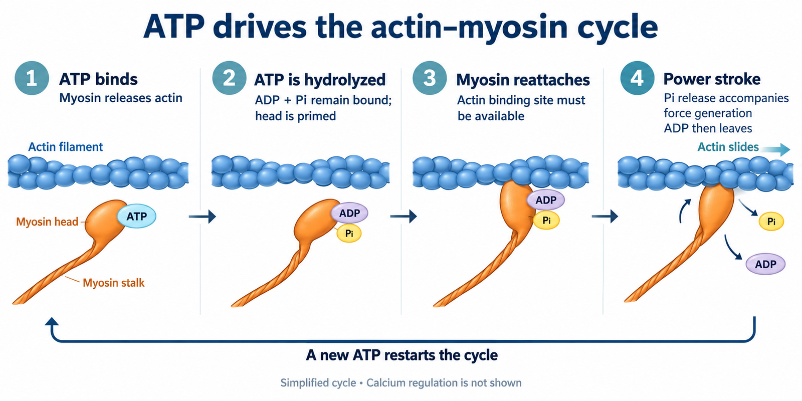
- A new ATP molecule binds to myosin. Myosin’s affinity for actin decreases, and it detaches. Notice that ATP binding causes this release.
- Myosin hydrolyzes ATP into ADP and Pi. Both products remain attached, while the myosin head changes into a configuration ready for its next stroke.
- Myosin binds to a new position on actin. The binding site must be accessible. Calcium helps regulate this accessibility, as explained below.
- A structural change associated with Pi release generates force. The myosin lever arm moves and transmits force to actin. This is the power stroke. ADP subsequently leaves as well.
- Another ATP binds, allowing detachment again. Many myosin heads repeat this cycle.
This is a simplified account of the basic mechanism. The essential distinctions are detachment with ATP binding, preparation following hydrolysis, and force generation associated with product release. These should not be collapsed into a single instant in which “ATP breaks down and the muscle gets shorter.” [2, 3]
Relaxation also requires ATP
When an electrical signal reaches a muscle cell, calcium ions, Ca²⁺, are released from the sarcoplasmic reticulum, a membrane-bound calcium store inside the cell.
Calcium binds troponin, a regulatory protein on the thin filament. This changes the position of tropomyosin, which otherwise covers myosin-binding sites on actin. Myosin can then attach. Calcium does not pull the filaments itself; it enables cross-bridge activity.
After the signal ends, calcium in the cytosol must fall for relaxation to occur. SERCA, the sarcoplasmic reticulum calcium pump, hydrolyzes ATP to move calcium back into storage. As calcium dissociates from troponin, the arrangement that blocks binding sites on actin is restored.
ATP therefore has at least two important roles:
- In the cross-bridge cycle, it allows myosin to detach from actin and prepare for another stroke.
- By powering calcium pumping, it removes the signal that permits contraction and restores the conditions for relaxation.
One myosin head detaching is not the same event as an entire muscle relaxing. If ATP becomes profoundly depleted, cross-bridges may instead become unable to detach. That extreme state should not be equated with ordinary exercise fatigue. [2, 3]
References
[1] OpenStax: ATP—Adenosine Triphosphate; Cooper: The Cell—Metabolic Energy.
[2] OpenStax: Muscle Fiber Contraction and Relaxation.
[3] Kuo and Ehrlich: Signaling in Muscle Contraction (2015).
Korean original reviewed for medical accuracy by S. Chang, KMD.
This article is for educational purposes and is not individual medical advice.
Written by: MedMaru Editorial Team
Reviewed for medical accuracy by:
S. Chang, KMD