Why Do Nerve Signals Travel in Only One Direction Along an Axon?
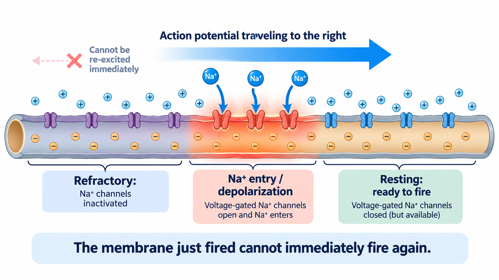
A nerve signal travels one way along an axon — the long, thin fiber a nerve cell uses to send messages — because the stretch of membrane it has just passed through is briefly unable to fire again. That short recovery window is called the refractory period. With the membrane behind it temporarily out of action, the signal has only one place to go: forward, into membrane that hasn’t fired yet.
How an action potential moves forward along the axon
The signal itself is an action potential, a brief spike in the voltage across the nerve cell’s membrane. It begins when voltage-gated sodium channels open. These are tiny pores that respond to a change in voltage by letting positively charged sodium ions flow into the cell. The inflow makes the inside of the membrane more positive, and that change spreads a short distance along the fiber, where it opens the next set of sodium channels and triggers the same spike again.
So no single packet of ions races from one end of the axon to the other. Each patch of membrane re-creates the spike in the patch beside it, much like a row of dominoes in which each piece knocks over the next. The comparison has a limit: a fallen domino stays down, while axon membrane resets itself shortly afterward and can fire again.
The refractory period: why the membrane behind the signal can’t fire
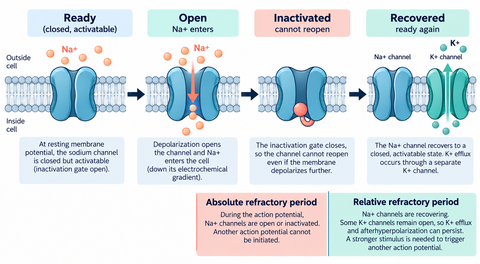
The same voltage change that opens the sodium channels also shuts down the membrane the signal has just left, in two ways. First, the sodium channels become inactivated. They close through a second gate, and in that state they cannot reopen no matter how strong the stimulus is. This stretch of time is the absolute refractory period.
Second, potassium channels open more slowly than sodium channels and stay open a little longer than needed. Potassium ions keep flowing out of the cell, and the membrane voltage briefly dips below its normal resting level, a dip called hyperpolarization. While it lasts, firing is possible again but harder, which is why this phase is often called the relative refractory period.
Put together, the membrane behind the signal is either unable to fire or reluctant to fire, while the membrane ahead is untouched and ready. That imbalance gives the signal its direction and explains why it never turns around and heads back toward the point where it began.
The refractory period has a second consequence. Because each patch of membrane needs time to recover, it also caps how many action potentials a neuron can fire per second. Different types of neurons carry different types and densities of ion channels, so their top firing rates differ as well.
Where the signal starts sets its direction
In a motor neuron, which carries commands from the spinal cord to a muscle, the action potential begins at the axon hillock, right next to the cell body, and travels outward along the axon toward the muscle. In a sensory neuron, the signal begins at the opposite end, at the nerve ending in the skin or an organ. When a fingertip is pricked, for example, the signal starts in the fingertip and travels inward toward the spinal cord and brain.
The two signals move in opposite directions, but they follow exactly the same rule. In both cases, the membrane the signal has just passed through is refractory, so the signal keeps moving away from where it started. The refractory period doesn’t send signals toward any particular destination. It only stops them from turning back, so the starting point decides the direction.
Antidromic conduction: when a signal travels the “wrong” way
If an axon is stimulated in the middle, where nothing has just fired, the membrane on both sides is ready, and the new signal spreads away from that point in both directions. The part moving in the nerve’s usual direction is called orthodromic conduction. The part moving the opposite way is called antidromic conduction.
This is not a malfunction, and clinicians use it on purpose. In nerve conduction studies, a test that measures how well nerves carry signals, sensory nerves are commonly stimulated antidromically: the electrode sends the signal against the nerve’s natural direction, and the response is recorded farther along the nerve. The nerve being assessed is the same either way. What changes is where the stimulating and recording electrodes sit.
Myelin speeds the signal but doesn’t change the rule
Many axons are wrapped in myelin, a fatty sheath that insulates the fiber. The sheath has regular gaps called nodes of Ranvier, and the signal leaps from node to node, which makes it travel much faster. Each node still generates its own action potential, so a recovery period still trails behind the signal. Myelin changes how fast the signal moves, not the reason it moves one way.
This article explains normal nerve physiology for learning purposes and is not medical advice. Persistent numbness, tingling, or weakness should be evaluated by a clinician.
References
- Purves D, Augustine GJ, Fitzpatrick D, et al., editors. “The Refractory Period.” Neuroscience. 2nd ed. Sunderland (MA): Sinauer Associates; 2001. NCBI Bookshelf NBK11146. https://www.ncbi.nlm.nih.gov/books/NBK11146/
- Grider MH, Jessu R, Kabir R. “Physiology, Action Potential.” StatPearls. Updated May 8, 2023. NCBI Bookshelf NBK538143. https://www.ncbi.nlm.nih.gov/books/NBK538143/
- Ramani PK, Lui F, Arya K. “Nerve Conduction Studies and Electromyography.” StatPearls. Updated February 10, 2025. NCBI Bookshelf NBK611987. https://www.ncbi.nlm.nih.gov/books/NBK611987/
Written by: MedMaru Editorial Team
Reviewed for medical accuracy by:
S. Chang, KMD