Amygdala: Location, Function, and Anatomy
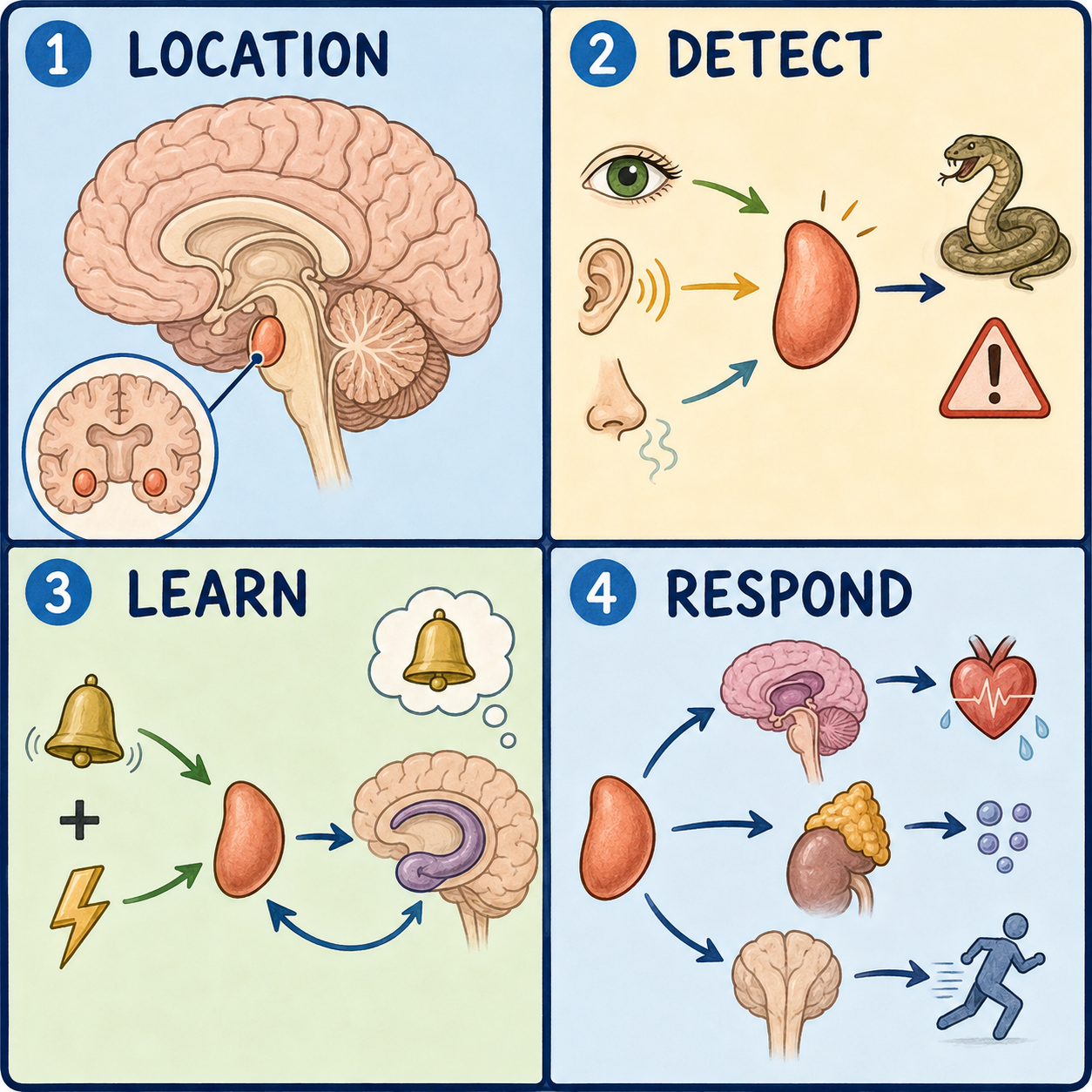
The amygdala is a small, almond-shaped cluster of neurons buried deep in each temporal lobe of the brain. Its best-known job is to notice possible danger quickly, often before you are consciously aware of it, and to help tag emotionally significant events, especially frightening or exciting ones, so they are more likely to be remembered. It works closely with several other brain regions, and despite its popular reputation, it is not a single “fear switch” that acts alone.
Not one structure, but a cluster
The name comes from the Greek word for almond, describing its general shape. Under the microscope, though, the amygdala isn’t a uniform blob. Researchers count roughly a dozen smaller clusters of neurons, called nuclei, organized into a few functional groups: the basolateral group, the cortical or corticomedial group, the central nucleus, and an extended region that connects to a structure called the bed nucleus of the stria terminalis. This extended amygdala is involved in longer-lasting stress and anxiety responses.
Each group handles a different part of the job. Some sort incoming sensory signals, some compare them against past experience, and some trigger an alarm response in the rest of the brain and body. Think of a smoke detector that also checks its records of past fires before sounding. Like a real smoke detector, the system is imperfect: the amygdala can flag something as threatening even when it turns out to be harmless.
Where it sits
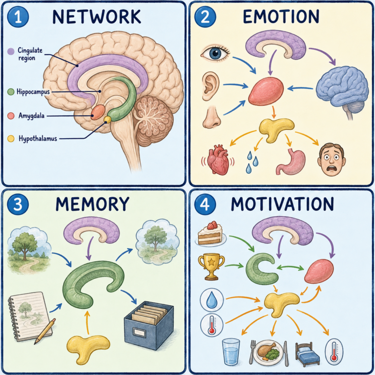
Each amygdala lies deep in the front part of a temporal lobe, just in front of the hippocampus and near the tip of the temporal horn of the lateral ventricle. This position puts it at a crossroads between incoming sensory information and the brain’s memory and stress-response systems.
Its output travels mainly along two pathways, the stria terminalis and the ventral amygdalofugal pathway, toward the hypothalamus, the septal area, and the brainstem. That wiring is how a perceived threat can trigger fast physical changes, like a racing heart, before you have consciously registered what caused it.
Fear, memory, and emotional tagging
The amygdala’s central role is in processing fear and other strong emotions, and in strengthening memories tied to them. When something seems dangerous or highly significant, the amygdala helps recruit the body’s stress response and works with the hippocampus to make the memory more vivid and durable. That is part of why frightening or emotionally intense experiences tend to be remembered more clearly than ordinary ones. It isn’t only about fear, though. The amygdala is involved more broadly in tagging emotionally significant experiences, including some positive or socially important ones. (For the wider picture, see how the brain produces fear and how the brain creates emotions.)
Research associated with the neuroscientist Joseph LeDoux proposed that the brain handles potential threats along two general routes: a faster, cruder pathway that can trigger a defensive reaction before the details are processed, and a slower pathway involving more detailed evaluation by the cortex. The framework is widely referenced, though the amygdala is only one part of a broader circuit, and researchers continue to refine how these pathways interact.
It also helps to separate two things that often get blurred together. The amygdala’s role in triggering measurable defensive behaviors, such as a startle response or a spike in heart rate, is not the same as the conscious feeling of fear or anxiety, which involves additional cortical processing. The distinction matters clinically, because a treatment that reduces one does not automatically resolve the other.
This is also why the popular phrase “amygdala hijack” is misleading. It is a popular expression, not a formal medical or neuroscience term, usually used for an intense emotional reaction that seems to override rational thought. In reality, the amygdala is one hub in a network that includes the hippocampus, prefrontal cortex, and hypothalamus, and its output is continuously shaped by input from those regions rather than acting as an independent trigger.
When the amygdala is affected
Because it sits within the temporal lobe, the amygdala can be affected by conditions involving that region. In temporal lobe epilepsy, seizures sometimes start near or involve the amygdala and hippocampus, and severe, treatment-resistant cases have occasionally been treated surgically by removing part of both structures. Some studies have also found differences in amygdala and hippocampal volume in people with certain mood and stress-related conditions. These findings reflect associations rather than a single simple cause, and volume differences alone don’t diagnose anything.
Damage to both amygdalae is rare. It typically results from specific circumstances, such as certain surgeries for severe epilepsy or rare genetic disorders. A dramatic illustration is Klüver-Bucy syndrome, which can follow damage to both temporal lobes, including both amygdalae. It combines reduced fear, indiscriminate eating, altered sexual behavior, and a tendency to explore objects by mouth. The syndrome is uncommon, but it shows how central this structure is to normal fear and emotional regulation.
This article is educational background, not a diagnostic guide. New seizures, or persistent anxiety or fear that interferes with daily life, should be discussed with a clinician.
Related: What Is the Cerebral Cortex? · What Is the Brain? · What Is the Cerebrum?
References
- StatPearls. “Neuroanatomy, Amygdala.” NCBI Bookshelf — https://www.ncbi.nlm.nih.gov/books/NBK537102/
- Kenhub. “Anatomy of the Amygdaloid Complex.” — https://www.kenhub.com/en/library/anatomy/anatomy-of-amygdaloidal-complex
- Cleveland Clinic. “Amygdala.” — https://my.clevelandclinic.org/health/body/24894-amygdala
- National Institute of Mental Health. “Circuitry for Fearful Feelings, Behavior Untangled in Anxiety Disorders.” Science Update, 2016 — https://www.nimh.nih.gov/news/science-updates/2016/circuitry-for-fearful-feelings-behavior-untangled-in-anxiety-disorders
- National Organization for Rare Disorders (NORD). “Kluver-Bucy Syndrome.” — https://rarediseases.org/rare-diseases/kluver-bucy-syndrome/
Written by: MedMaru Editorial Team
Reviewed for medical accuracy by:
S. Chang, KMD