Insular Cortex : What It Does and Why It’s Hidden in the Brain
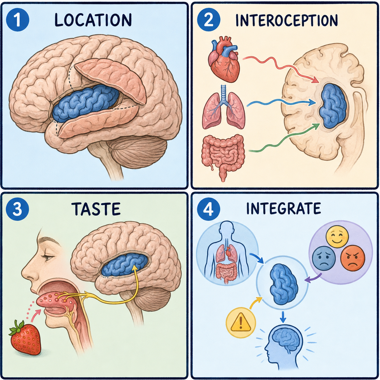
The insular cortex, or insula, is a part of the brain’s outer layer that you cannot see from the outside. The outer layer, the cortex, is the wrinkled surface where most thinking and sensing begins. The insula is tucked deep inside a groove on the side of the brain, out of view under other tissue. Three overlapping flaps of surrounding cortex, called opercula, from the Latin for lids, fold over it like a hand cupped over a small object. The insula forms the floor of the lateral sulcus, also called the Sylvian fissure, the deep groove that separates the frontal and parietal lobes above from the temporal lobe below. There is one insula in each hemisphere.
Why a piece of cortex ended up hidden
The insula is sometimes taught as a fifth lobe of the cerebral cortex, sitting where the frontal, parietal, and temporal lobes meet. That label is a teaching convenience rather than a strict anatomical rule. Unlike the four classic lobes, the insula has no direct opening to the brain’s outer surface. It is completely enclosed.
Why would the brain fold a piece of cortex inward instead of leaving it exposed? During fetal development, the surrounding frontal, parietal, and temporal cortex grows faster and farther than the insula. The faster-growing tissue curls over and around it and seals it inside a fissure. The insula is not lost or vestigial. Its spot at the junction of three lobes fits its role in blending signals arriving from all three.
Imagine folding a sheet of paper down over a coin on a table, then folding a second sheet up from underneath to meet it. The coin is still there and still attached to the table, but you have to peel the paper back to see it. The analogy has a limit. Real cortex is one continuous, wrinkled sheet of neurons, not separate paper flaps, so the insula is physically continuous with the tissue folded over it rather than a separate object trapped underneath. That is also why it only becomes visible once the lateral sulcus is opened or the opercula are gently retracted.
The lids, the borders, and the neighbors
The three lids each have a name. The frontal, or fronto-orbital, operculum covers the insula from the front. The frontoparietal operculum covers it from above and is formed by the lower parts of the precentral, postcentral, and supramarginal gyri. The temporal operculum covers it from below and is formed mainly by the superior temporal gyrus. A neurosurgeon who needs to reach the insula has to work through or around these opercula. That is one reason insular surgery is technically demanding: the target sits beneath cortex that itself handles movement, sensation, and language.
A groove called the circular sulcus, or peri-insular sulcus, runs around almost the whole edge of the insula and separates it from the opercula. At its front-lower corner, the insula narrows to a point called the limen insulae, the threshold of the insula. There its surface blends into the medial temporal lobe near the anterior perforated substance, a small patch of brain surface where blood vessels enter.
Inside, the central insular sulcus splits the insula into front and back halves, much as the central sulcus divides the frontal and parietal lobes on the outer surface. The front half has three short gyri separated by two precentral insular sulci. The back half has two long gyri separated by one postcentral insular sulcus. The split matters because the two halves connect to different parts of the brain.
Moving inward from the insula toward the middle of the brain, the layers run in a fixed order. First comes the extreme capsule, a thin band of white matter. Next is the claustrum, a thin sheet of gray matter. Then comes the external capsule, another band of white matter, and finally the putamen, part of the basal ganglia. This puts the insula right next to deep movement-control structures. It is one reason insular strokes or tumors can sometimes cause motor symptoms, even though the insula is not mainly a movement-control region.
Blood reaches the insula through short perforating branches of the M2, or insular, segment of the middle cerebral artery, the MCA. Because these branches are short and numerous, the insula is vulnerable when a stroke affects MCA territory.
A hub with front and back wiring
A common misconception is that the insula, being small and hidden, doesn’t matter much. Its hidden location says nothing about its importance. The insula links to the surrounding frontal, parietal, and temporal cortex. It links to limbic structures, including the olfactory system, the anterior cingulate cortex, the amygdala, the hippocampus, and the thalamus. It also links to the basal ganglia.
Those connections are not spread evenly. A 2025 narrative review reports that the front of the insula connects mainly with the anterior cingulate cortex and with frontal, orbitofrontal, and anterior temporal areas. The back of the insula connects mainly with posterior temporal, parietal, and sensorimotor areas. This front-versus-back pattern is the anatomical basis for linking the anterior insula with emotional and internal-state processing, and the posterior insula with sensory and motor processing. Internal-state processing includes interoception, the sense of the body’s own condition, such as heartbeat, hunger, or breathlessness. Its link to the anterior insula is still an area of active research.
For the same reason, it is a mistake to say the insula does just one thing, whether that is taste, emotion, or pain. No single function is established. The anatomy shows connections to sensory, motor, limbic, and cognitive-control regions all at once.
Why insular damage looks so varied
Damage to the insula rarely produces one clean symptom, because it borders motor, sensory, language, and limbic territory at the same time. In insular strokes, reported findings include weakness or clumsiness on one side, slurred speech, called dysarthria, and language difficulty, called aphasia. Hearing or balance disturbances, altered taste, and behavioral or psychiatric changes have also been reported.
Seizures that start in the insula have been reported to produce sensory or taste-related warning sensations, autonomic disturbances, swallowing difficulty, a feeling of throat tightening, unusual movements, and dizziness or vertigo. Gliomas, a type of brain tumor, also occur here at a notable rate: an estimated 10 to 25 percent of intracranial gliomas involve the insula. Surgery in this area is high-risk because the insula lies directly beside the M2 segment of the MCA, under cortex responsible for movement and language.
This article describes anatomy for learning purposes. It does not cover treatment decisions, does not estimate individual prognosis, and does not replace evaluation by a neurologist or neurosurgeon.
Related: What Is the Cerebral Cortex? · What Is the Brain? · What Is the Cerebrum?
References
- StatPearls. “Insular Cortex.” NCBI Bookshelf, NBK570606 — https://www.ncbi.nlm.nih.gov/books/NBK570606/
- StatPearls. “Neuroanatomy, Sylvian Fissure.” NCBI Bookshelf, NBK574552 — https://www.ncbi.nlm.nih.gov/books/NBK574552/
- “Insular Cortex — Biology and Its Role in Psychiatric Disorders: A Narrative Review.” PMC, 2025, PMC12384716 — https://pmc.ncbi.nlm.nih.gov/articles/PMC12384716/
Written by: MedMaru Editorial Team
Reviewed for medical accuracy by:
S. Chang, KMD