What Is the Brain? Parts, Functions, and How It Works
If you could hold an adult human brain, it would weigh about 1.3 to 1.4 kilograms, roughly the weight of a large bag of sugar. It is soft, wrinkled, and mostly water. Nothing about it looks like a machine. Yet this organ decides where your eyes point, keeps you breathing through the night, and holds every memory you have.
So what exactly is it? The simplest definition is this: the brain is the part of the nervous system inside the skull that receives information from the body and the outside world, makes sense of it, and sends out the signals that control what the body does. Anatomists divide it into four main parts, the cerebrum, the diencephalon, the brainstem, and the cerebellum, and almost everything it does is a variation on one loop: take information in, work out what it means, and turn it into action. This article walks through where the brain sits, what it is built from, what each part does, and how the parts work together.
Where the brain fits in the nervous system
The brain does not work alone. Together with the spinal cord, it forms the central nervous system, the body’s main processing center. Everything outside the brain and spinal cord, including the nerves running to your skin, muscles, and organs, belongs to the peripheral nervous system.
The two systems form a loop. Peripheral nerves carry information in, the brain and spinal cord interpret it, and peripheral nerves carry commands back out. The brain sits at the top of that loop, which is why damage to it can affect almost anything the body does. For the bigger picture, see what the nervous system is.
How the brain is protected
Brain tissue is delicate, so the body surrounds it with several layers of defense. The skull is the obvious one. Beneath it lie the meninges, three membranes that wrap the brain and spinal cord. Between two of those layers flows cerebrospinal fluid, a clear liquid that cushions the brain so it effectively floats rather than resting its full weight against bone.
The last layer of protection is chemical rather than physical. The blood vessels in the brain are lined with cells packed unusually tightly together, forming the blood-brain barrier. It lets oxygen and nutrients through while keeping out many substances in the blood that could disturb the finely balanced environment neurons need.
The four main parts of the brain
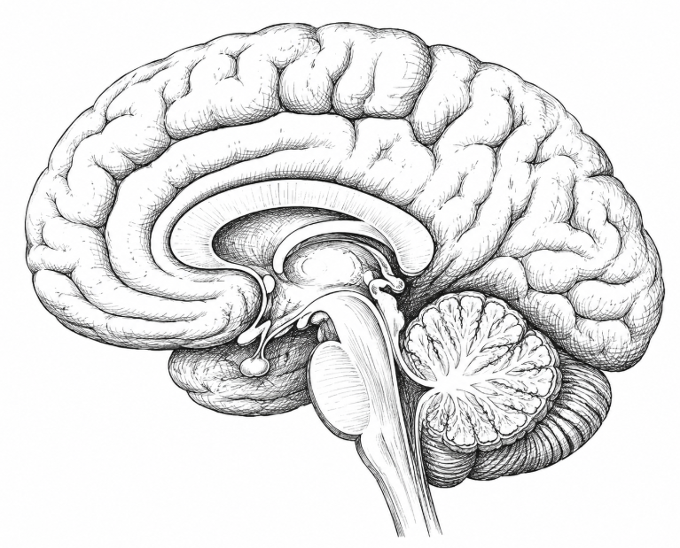
The easiest way to picture the four main parts is from the midline, as if the brain had been sliced down the middle from front to back.
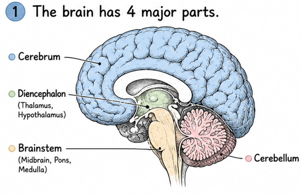
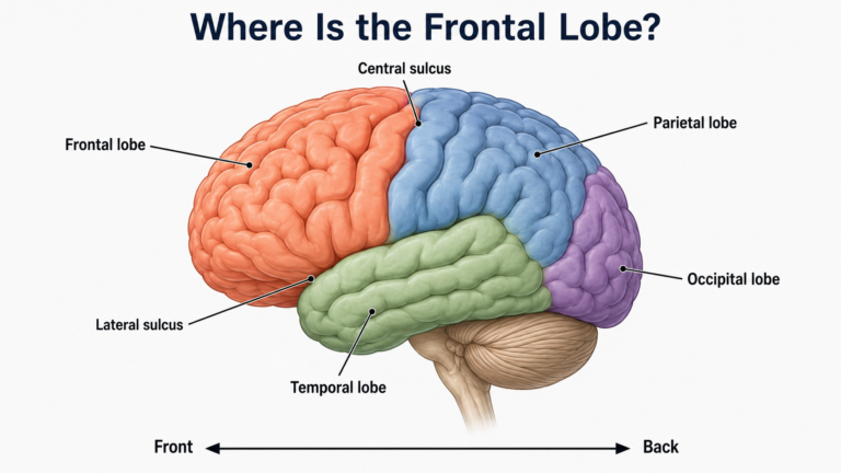
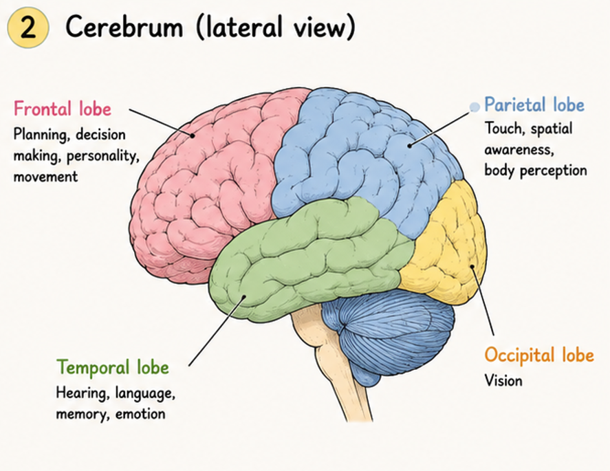
The cerebrum is the large, folded part on top. It accounts for roughly 80 to 85 percent of the brain’s volume and is divided into left and right hemispheres, joined by a thick band of fibers called the corpus callosum. Its outer layer, the cerebral cortex, is folded into ridges and grooves and handles much of perception, language, planning, and voluntary movement. The cortex is usually mapped into four lobes: frontal, parietal, temporal, and occipital.
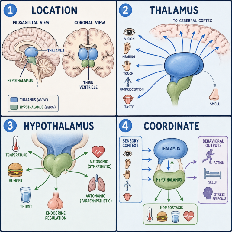
Tucked beneath the cerebrum is the diencephalon. Its two best-known structures are the thalamus, which relays nearly all incoming sensory information to the cortex (smell is the exception), and the hypothalamus, which helps regulate body temperature, hunger, thirst, sleep, and hormones. The thalamus is more than a relay, though; it also takes part in motor circuits, attention, and consciousness.
The brainstem runs down from there to the spinal cord and is made up of the midbrain, pons, and medulla oblongata. It handles functions you never think about, such as breathing, heart rate, blood pressure, and swallowing, it contains many of the nuclei of the cranial nerves, and it helps keep you awake and alert. Because so much depends on it, severe brainstem injury is often life-threatening.
Behind the brainstem sits the cerebellum. It makes up only about 10 percent of the brain’s volume, yet it contains more than half of all the brain’s neurons. It fine-tunes movement, balance, posture, and timing, and it is essential for learning motor skills. It does not start movements; it compares what you intended with what your body is actually doing and corrects the difference.
These divisions are useful labels, but the parts are tightly wired to one another, as the rest of this article shows.
What the brain is made of
At the cellular level, the brain is built from two broad kinds of cells. Neurons carry signals, and the adult human brain contains around 86 billion of them. Glial cells support, insulate, and protect neurons, and they are present in roughly similar numbers. That finding overturned an older, widely repeated claim that glia outnumber neurons ten to one.
Those cells are arranged in two kinds of tissue you can see with the naked eye. Gray matter is rich in neuron cell bodies and forms the cortex and clusters deeper inside the brain. White matter is made mostly of long nerve fibers wrapped in myelin, a fatty coating that speeds up signals and gives the tissue its pale color. Gray matter is broadly where processing happens, and white matter is the wiring that connects one area to another.
What the brain does
Picture the first minute of an ordinary morning. The alarm goes off, you reach over to silence it, you notice the room is cold, you remember you have a meeting at nine, and you feel a small wave of dread about it. None of that felt like work. Yet in those few seconds your brain took in sound, located your hand in space, moved it, read your body temperature, pulled up a memory, and attached an emotion to it, all while keeping your heart beating and your lungs breathing.
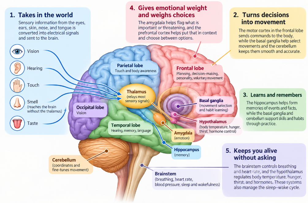
It takes in the world. Your senses do not send pictures or sounds to the brain. They send electrical signals. Receptors in the eyes, ears, skin, nose, and tongue convert light, pressure waves, touch, and chemicals into nerve impulses. Most of these signals pass first through the thalamus before reaching the areas of the cortex that specialize in each sense: vision mainly in the occipital lobe, hearing in the temporal lobe, and touch in the parietal lobe. The experience of seeing a red cup or hearing your name is something the brain builds from these signals, not something it simply receives. (More on that in how the brain processes sensory information.)
It turns decisions into movement. When you decide to pick up that cup, the command starts in the motor cortex, a strip of tissue toward the back of the frontal lobe. Signals travel down through the brainstem and spinal cord to the nerves that control your muscles. Each side of the motor cortex mainly controls the opposite side of the body, which is why damage to one side of the brain can weaken the other side. The cerebellum corrects the movement as it happens, and the basal ganglia help select which movement to make and suppress the ones you do not want. That cooperation is why reaching for a cup is smooth rather than jerky. (How the brain controls movement goes further.)
It learns and remembers. Learning is the brain changing itself. Each time you practice a skill or take in a new fact, the connections between neurons, called synapses, can grow stronger or weaker. This capacity to rewire with experience is called neuroplasticity, and it continues throughout life. Different kinds of memory lean on different systems. Remembering what happened yesterday depends heavily on the hippocampus, deep in the temporal lobe, while learning to ride a bike relies more on the basal ganglia and cerebellum. That split explains why someone can lose the ability to form new memories of events yet still pick up a new motor skill. (How the brain forms memories covers this in detail.)
It gives experience emotional weight and weighs choices. The amygdala helps flag what is threatening or important, which is why a frightening moment is often remembered more vividly than an ordinary one. The prefrontal cortex helps put those signals in context, hold goals in mind, and choose between options. Emotion and reasoning are not opposites here; decisions draw on both. (How the brain creates emotions and how the brain makes decisions explore each side.)
It keeps you alive without asking. Some of the brain’s most important work never reaches awareness. The brainstem controls breathing and helps regulate heart rate and blood pressure. The hypothalamus acts as a thermostat for body temperature, generates hunger and thirst, and directs much of the hormone system through the pituitary gland just beneath it. These systems also manage the daily cycle of sleep and wakefulness. This background regulation continues through sleep, anesthesia, and deep concentration. (See how the brain controls the autonomic nervous system.)
Why no single part does it alone
It is tempting to picture the brain as a set of rooms, each with one job. The real picture is closer to a network. Regions do specialize, but almost every everyday task recruits many of them at once. Picking up a cup draws on the cerebrum to plan the movement, the brainstem and spinal cord to carry the commands, the cerebellum to fine-tune them, and the diencephalon to relay sensation and keep track of the body’s internal state.
Speaking a single sentence is an even richer example. You need language areas to find the words and arrange them, the motor cortex and cerebellum to move your lips, tongue, and breath precisely, hearing areas to monitor your own voice, memory to recall what you mean, and attention to stay on topic. Damage to one region can therefore disrupt a function without erasing it, and other areas can sometimes partly take over with time and practice.
This constant traffic is expensive. The brain makes up only about 2 percent of an adult’s body weight but uses roughly 20 percent of the body’s energy at rest. Most of that energy goes into keeping its neurons ready to signal to one another.
How the brain works at the level of cells
Everything the brain does comes down to neurons signaling to one another. A neuron sends a brief electrical impulse, called an action potential, down its long fiber. When the impulse reaches the end, it triggers the release of chemical messengers called neurotransmitters into a tiny gap, the synapse, where they act on the next cell.
One neuron can connect with thousands of others, and the patterns of those connections are what matter. They form circuits and networks that let you recognize a face, recall a word, or catch a falling glass. Those connections are also not fixed. They strengthen, weaken, and reorganize with experience, which is how the brain learns. (How neurons communicate explains the signaling step by step.)
Two ideas about the brain worth unlearning
The first is the claim that we use only 10 percent of our brains. Brain imaging and studies of brain injury show otherwise: every region has a role, and even small areas of damage can cause noticeable problems. Not every region is highly active at every moment, but over the course of a day, virtually all of the brain is put to work.
The second is that the adult brain can never make new neurons. For most regions, lost neurons are indeed not replaced. But research since the late 1990s has found evidence that new neurons form in at least one area of the adult human brain, the hippocampus, which is important for memory. How many are produced, and how much they matter for everyday function, is still being actively studied.
Why this map matters in medicine
Knowing the four main parts helps explain why different neurological problems produce different symptoms. A stroke affecting the frontal lobe may impair speech or movement. Damage to the cerebellum may cause poor coordination. Injury to the brainstem can affect breathing and consciousness. Disorders of the hypothalamus may disrupt body temperature or hormone regulation. Real injuries rarely respect these boundaries neatly, which is why diagnosis relies on a clinical examination and, often, brain imaging.
This article is for general education and does not replace advice from a qualified clinician. Sudden weakness, numbness, trouble speaking, loss of coordination, or reduced alertness needs emergency medical care.
References
- Purves D, Augustine GJ, Fitzpatrick D, et al., editors. Neuroscience. 2nd ed. Sinauer Associates; 2001. NCBI Bookshelf — https://www.ncbi.nlm.nih.gov/books/NBK10799/
- National Institute of Neurological Disorders and Stroke. Brain Basics: Know Your Brain. National Institutes of Health. — https://www.ninds.nih.gov/health-information/public-education/brain-basics/brain-basics-know-your-brain
- Azevedo FAC, Carvalho LRB, Grinberg LT, et al. Equal numbers of neuronal and nonneuronal cells make the human brain an isometrically scaled-up primate brain. Journal of Comparative Neurology. 2009;513(5):532-541. doi:10.1002/cne.21974
- Daneman R, Prat A. The blood-brain barrier. Cold Spring Harbor Perspectives in Biology. 2015;7(1):a020412. doi:10.1101/cshperspect.a020412
- Eriksson PS, Perfilieva E, Björk-Eriksson T, et al. Neurogenesis in the adult human hippocampus. Nature Medicine. 1998;4(11):1313-1317. doi:10.1038/3305
- Disouky A, Sanborn MA, Sabitha KR, et al. Human hippocampal neurogenesis in adulthood, ageing and Alzheimer’s disease. Nature. 2026;652(8112):1264–1273. doi:10.1038/s41586-026-10169-4. https://www.nature.com/articles/s41586-026-10169-4
- Standring S, ed. Gray’s Anatomy: The Anatomical Basis of Clinical Practice. 42nd ed. Elsevier; 2020.
- National Institutes of Health. Information about the Brain. NIH Curriculum Supplement Series. NCBI Bookshelf — https://www.ncbi.nlm.nih.gov/books/NBK20367/
- Bui T, Das JM. Neuroanatomy, Cerebral Hemisphere. StatPearls. NCBI Bookshelf — https://www.ncbi.nlm.nih.gov/books/NBK549789/
- Basinger H, Hogg JP. Neuroanatomy, Brainstem. StatPearls. Updated July 4, 2023.
- Jimsheleishvili S, Dididze M. Neuroanatomy, Cerebellum. StatPearls. NCBI Bookshelf.
Written by: MedMaru Editorial Team
Reviewed for medical accuracy by:
S. Chang, KMD