What Are the Meninges? 3 Layers of the Brain Explained
The meninges are three thin sheets of connective tissue that wrap the brain and spinal cord. They hold the brain in place, cushion it in fluid, and carry blood vessels across its surface.
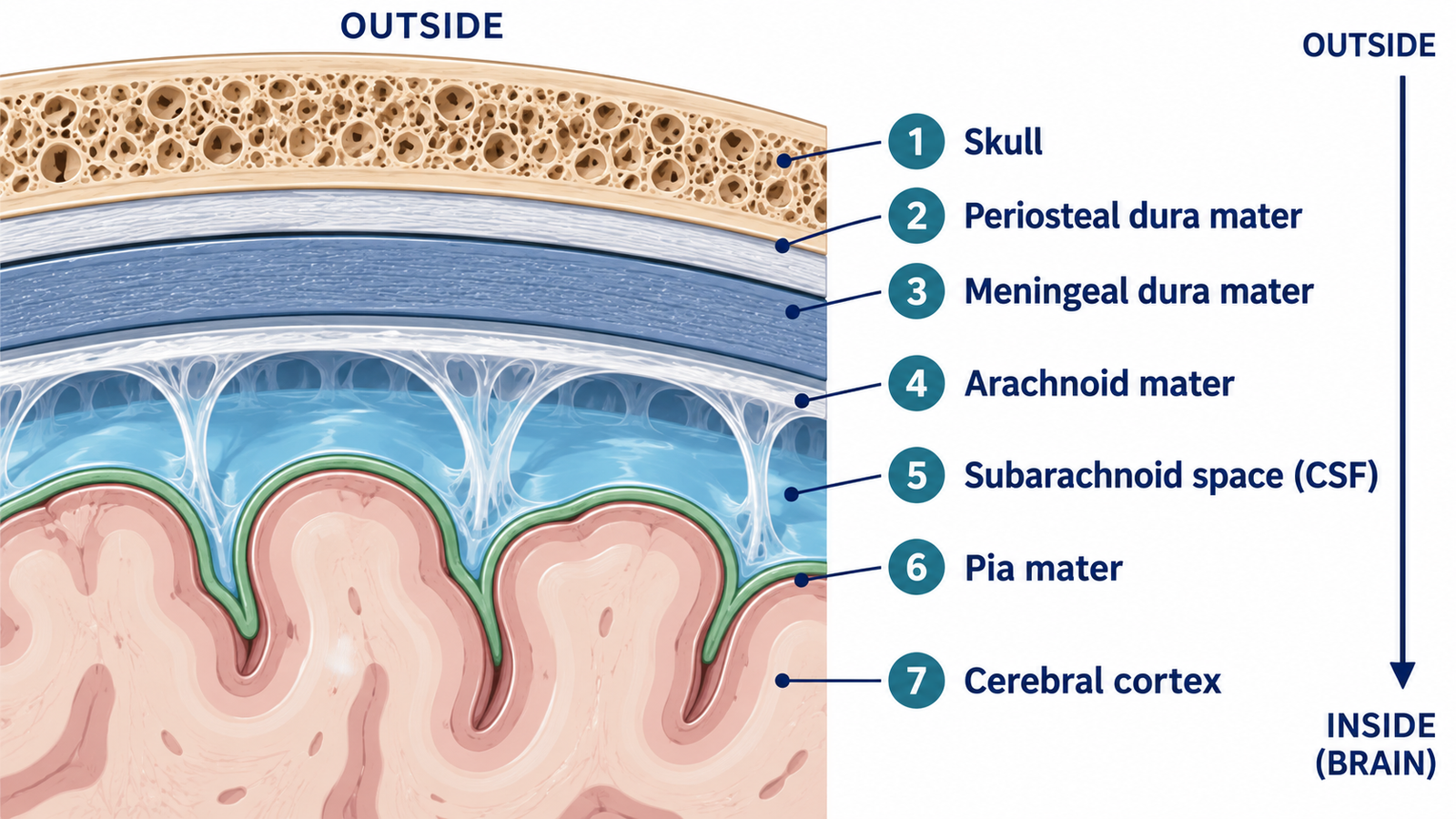
From the skull inward, the three layers are the dura mater, the arachnoid mater, and the pia mater. The dura is the tough outer sheet. The arachnoid is a delicate middle web. The pia is a thin film attached to the brain itself. Between the arachnoid and the pia lies the subarachnoid space, which is filled with cerebrospinal fluid, the clear liquid the brain floats in. When these layers become inflamed, usually because of infection, the result is meningitis.
Three layers, always in the same order
The word meninges (say: meh-NIN-jeez) simply means “membranes.” In anatomy, it refers to the three layers that cover the entire central nervous system, brain and spinal cord together. Their order never changes. From the surface inward, it is dura, arachnoid, pia.
Think of the packaging around a fragile object in a shipping box. There is a stiff outer carton, a loose web of padding, and a film clinging to the object itself. The comparison stops there. Packaging does nothing else, while the meninges carry blood, drain fluid, and sense pain.
It also helps to separate two ideas that are easy to blur. The skull is bone. The meninges are soft tissue lying beneath it.
The dura mater: a tough outer layer with several jobs
Dura mater is Latin for “tough mother,” and the name fits. It is the thick, leathery outermost layer. Inside the skull it is really two sheets pressed together. The periosteal layer lies against the bone, and the meningeal layer faces the brain.
In some places the two sheets separate and fold inward, forming stiff partitions. The falx cerebri is a sickle-shaped sheet that hangs down between the two cerebral hemispheres. The tentorium cerebelli is a U-shaped shelf that separates the cerebellum below from the occipital lobes above. These folds limit how far the brain can shift when the head moves or turns, much like cardboard dividers in a box of glassware. Unlike those dividers, the folds are continuous with the outer covering and carry blood inside them.
That blood travels in channels called dural venous sinuses, which form where the two dural sheets part. They are the brain’s main route for venous drainage. So a blockage here is not a problem in one small vein. It obstructs a major exit route for blood leaving the brain. A clot in a dural venous sinus can also block the reabsorption of cerebrospinal fluid, which drains into these same sinuses, and this raises the pressure inside the skull.
The dura also explains something about headaches. Brain tissue itself is insensitive to pain. Parts of the dura, the large venous sinuses, and the large arteries at the base of the brain are pain-sensitive. The meninges, by contrast with brain tissue, have a nerve supply. It comes mainly from the vagus and trigeminal nerves, with some cervical spinal nerves contributing to a lesser degree. This is why pain can seem to come from “inside the head” even though brain tissue itself does not register it.
The arachnoid mater and the fluid beneath it
Arachnoid comes from the Greek word for spider. The fine strands that cross beneath this layer look like a web. The arachnoid has no blood vessels of its own. Its importance lies in the space directly under it.
The subarachnoid space sits between the arachnoid and the pia, and it is filled with cerebrospinal fluid. This is where the brain’s cushioning happens. At any given time, about 150 mL of cerebrospinal fluid circulates. Roughly 25 mL is inside the ventricles, and the rest is in the subarachnoid space. The choroid plexus produces it at about 0.3 mL per minute. So most cerebrospinal fluid lies outside the brain, not inside it.
A brain in fluid is like an egg suspended in a jar of water rather than resting on a hard shelf. A sudden knock is spread out instead of landing on one point. The comparison has a limit, because cerebrospinal fluid also carries away waste and transports signaling molecules, which plain water in a jar does not do.
The fluid does not simply pool. Small projections of the arachnoid, called arachnoid granulations or arachnoid villi, push into the superior sagittal sinus, one of the dural venous sinuses. They are the main route by which cerebrospinal fluid is reabsorbed into the blood. On this main return path, fluid moves from the subarachnoid space, through the arachnoid granulations, into a dural venous sinus, and back to the general circulation. If absorption along this route is impaired, fluid can build up faster than it drains. The ventricles then widen, and pressure inside the skull can rise. This is one form of hydrocephalus, called communicating hydrocephalus, and it can follow bleeding into the subarachnoid space or inflammation such as meningitis.
The pia mater: the layer that follows every fold
Pia mater means “tender mother.” It is the innermost and thinnest layer. The arachnoid bridges across the brain’s grooves, but the pia clings directly to the surface of the brain and spinal cord. It dips into every sulcus and rides over every gyrus. Picture cling film pressed onto a walnut, with the arachnoid stretched loosely over the top like a sheet. The cling-film picture falls short in one way: the pia is living tissue, and blood vessels travel along it as they enter the brain.
In the spinal canal, the pia continues past the tapered end of the spinal cord and fuses into the filum terminale, which anchors the cord to the coccyx. Along the sides of the cord, the pia forms denticulate ligaments that anchor it to the spinal dura.
Only one of the “spaces” normally exists inside the skull
Because there are three layers, it is natural to assume there are three matching gaps between them. Inside the skull, that is not the case.
| Space | Location | Present normally? | Clinical association |
|---|---|---|---|
| Epidural (cranial) | Between skull and dura | No — potential space only | Epidural hematoma; classically middle meningeal artery tear with temporal bone fracture |
| Subdural | Between dura and arachnoid | No — potential space only | Subdural hematoma; classically bridging vein rupture |
| Subarachnoid | Between arachnoid and pia | Yes — filled with CSF | Subarachnoid hemorrhage; thunderclap headache |
| Epidural (spinal) | Between ligamentum flavum and dura | Yes — fat, veins, nerves, lymphatics | Site of epidural injection |
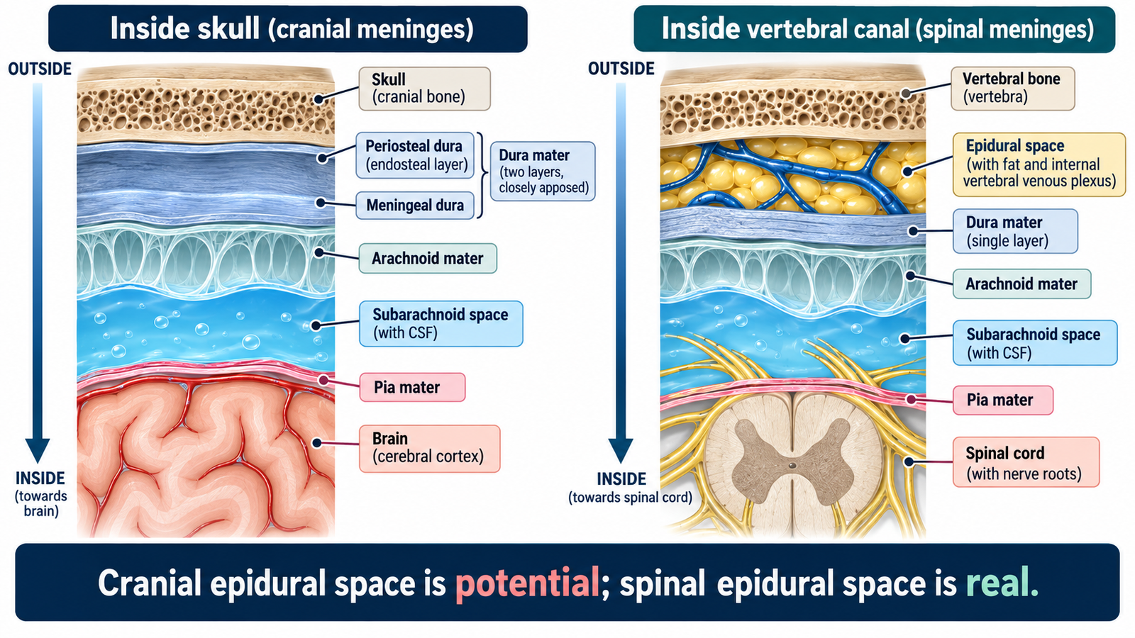
Inside the skull, the outer dura is firmly attached to the periosteum of the skull. As a result, the cranial epidural space typically exists only in disease, and the same is true of the subdural space. This has a practical meaning. An epidural or subdural bleed does not fill a pocket that was already there. It forces a space open.
The classic source of bleeding differs by location. An epidural hematoma is usually caused by rupture of the middle meningeal artery following a fracture of the temporal bone. A subdural hematoma usually results from rupture of the bridging veins. Bleeding into the subarachnoid space, which is already filled with fluid, has a hallmark symptom: a sudden thunderclap headache.
How the spinal meninges differ
The spine follows different rules. In the spinal canal, the epidural space is real and occupied. It contains fat, connective tissue, nerves, the internal vertebral venous plexus, and lymphatics. This is why an epidural injection can be given in the back but not in the head. The spinal dura is also simpler. It has only the meningeal layer, because the periosteal layer ends at the foramen magnum.
Why a lumbar puncture is done low in the back
Cerebrospinal fluid can be sampled from the lower back because the subarachnoid space extends below the end of the spinal cord. The cord tapers to an end called the conus medullaris. This usually lies around the first or second lumbar vertebra, on average at the middle third of L1, and it can sometimes be lower. Below it, the fluid-filled space continues as the lumbar cistern. In adults, the L3/L4 interspinous space is the preferred site for collecting cerebrospinal fluid safely.
Meningitis and when to seek care
The meninges surround the whole central nervous system without a break. For that reason, inflammation of these layers affects the entire system rather than one spot. The CDC defines meningitis as inflammation of the lining of the brain and spinal cord. Its causes include viral, bacterial, and fungal infections, parasites, amebas, injuries, cancer, and certain drugs.
Common symptoms are fever, headache, and a stiff neck. Nausea, vomiting, sensitivity to light, and confusion can also occur. CDC guidance is direct: anyone with symptoms of meningitis should see a healthcare provider right away. A sudden, severe headache that reaches maximum intensity within seconds, often described as the worst ever, is the classic description of subarachnoid hemorrhage and is an emergency.
This article describes normal anatomy for learning purposes. It is not a diagnosis, does not provide criteria for ruling any condition in or out, and does not replace assessment by a clinician.
References
- Neuroanatomy, Cranial Meninges. StatPearls. NCBI Bookshelf. https://www.ncbi.nlm.nih.gov/books/NBK539882/
- Anatomy, Back, Spinal Meninges. StatPearls. NCBI Bookshelf. https://www.ncbi.nlm.nih.gov/books/NBK547755/
- Anatomy, Head and Neck, Subarachnoid Space. StatPearls. NCBI Bookshelf. https://www.ncbi.nlm.nih.gov/books/NBK557521/
- Neuroanatomy, Conus Medullaris. StatPearls. NCBI Bookshelf. https://www.ncbi.nlm.nih.gov/books/NBK545227/
- Centers for Disease Control and Prevention. About Meningitis. Last reviewed September 9, 2025. https://www.cdc.gov/meningitis/about/index.html
- Headache. In: Walker HK, Hall WD, Hurst JW, eds. Clinical Methods: The History, Physical, and Laboratory Examinations. 3rd ed. NCBI Bookshelf. https://www.ncbi.nlm.nih.gov/books/NBK377/
- Cerebral Venous Sinus Thrombosis. StatPearls. NCBI Bookshelf. https://www.ncbi.nlm.nih.gov/books/NBK459315/
- Hydrocephalus. StatPearls. NCBI Bookshelf. https://www.ncbi.nlm.nih.gov/books/NBK560875/
- National Institute of Neurological Disorders and Stroke. Hydrocephalus. https://www.ninds.nih.gov/health-information/disorders/hydrocephalus
Written by: MedMaru Editorial Team
Reviewed for medical accuracy by:
S. Chang, KMD