What Is the Autonomic Nervous System? Functions and Divisions
Your heart keeps beating while you sleep. You keep breathing without thinking about it. Your stomach digests food, your pupils adjust to the light, and your blood pressure is constantly fine-tuned. All of this happens automatically because it is managed by the autonomic nervous system, or ANS. The autonomic nervous system is the division of the peripheral nervous system that automatically regulates heart muscle, smooth muscle, and glands. It maintains homeostasis, the body’s stable internal balance, by controlling heart rate, blood pressure, breathing, digestion, body temperature, and pupil size without conscious effort.
Automatic, but not on its own
The peripheral nervous system has two major functional divisions. The somatic nervous system supplies skeletal muscle and handles voluntary movement. The autonomic nervous system supplies smooth muscle, cardiac muscle, and glands, tissues that keep working whether you are awake or asleep. It operates largely outside conscious awareness.
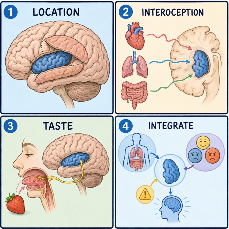
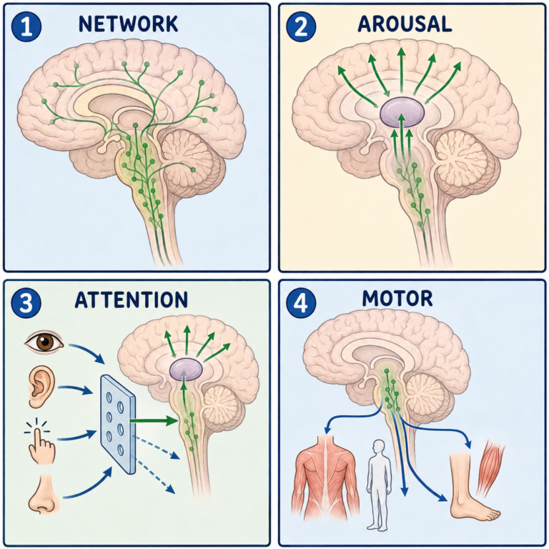
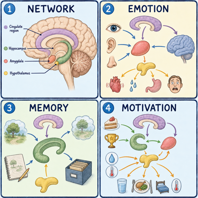
That doesn’t mean it works independently of the brain. Many autonomic reflexes happen automatically, but higher brain regions, including the hypothalamus, the brainstem, the insula, and the anterior cingulate cortex, play essential roles in regulating it. Think of a building’s automatic climate control. It runs on its own most of the time, but building managers can still adjust the settings. The analogy breaks down in that the brain’s oversight is continuous rather than occasional, but it shows how automatic and central control coexist.
A two-neuron relay
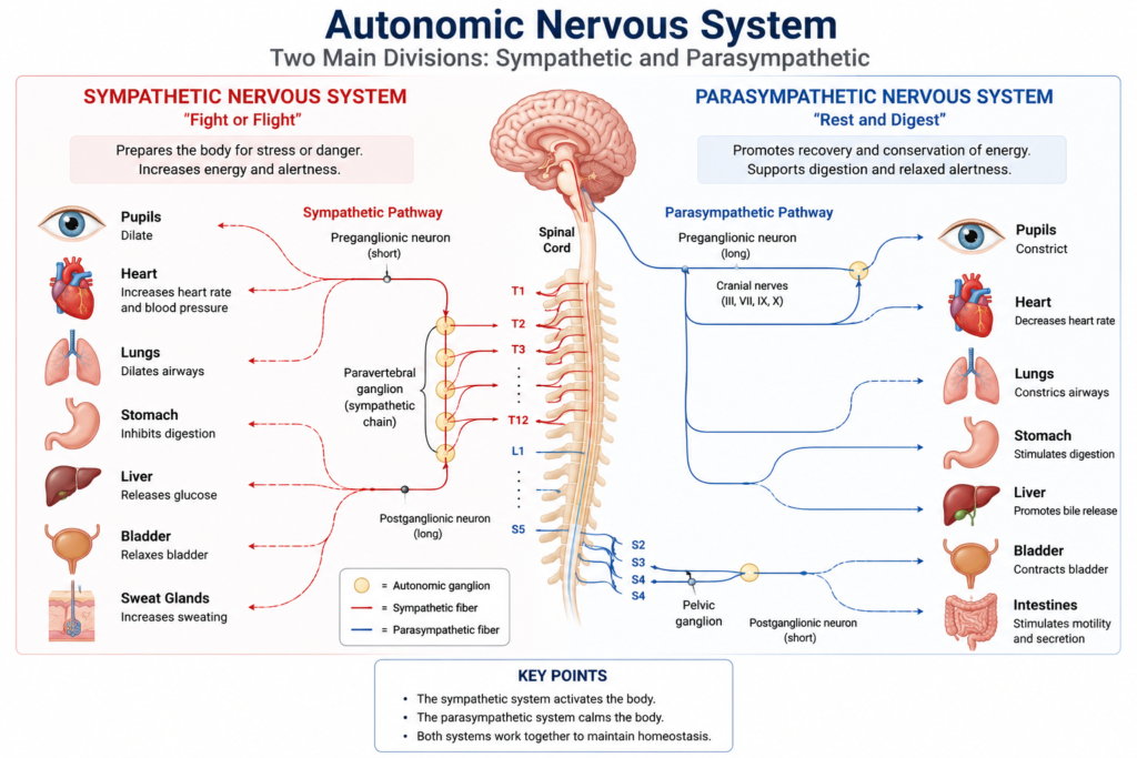
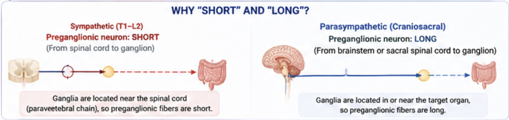
A key feature of the autonomic system is that its outgoing motor pathway uses two neurons rather than one. The first, the preganglionic neuron, starts in the brainstem or spinal cord. Its axon travels to an autonomic ganglion, a cluster of nerve cells outside the central nervous system, and connects there with the second neuron. The second, the postganglionic neuron, starts in the ganglion and sends its axon to the target organ, where it regulates muscle contraction, gland secretion, or heart activity.
This two-step design is one of the main things that sets the autonomic system apart from the somatic system, which uses a single motor neuron running from the central nervous system all the way to the muscle. The somatic motor neuron ends at a neuromuscular junction, while autonomic fibers end at a neuroeffector junction on their target tissue.
Three divisions working together
The autonomic nervous system has three functional divisions. The sympathetic nervous system prepares the body for increased physical or mental activity and is commonly linked to the fight-or-flight response. It raises heart rate and blood pressure, widens the pupils and airways, reduces digestive activity, and increases the availability of glucose, helping the body respond quickly to stress or danger.
The parasympathetic nervous system promotes recovery and energy conservation and is often called the rest-and-digest system. It slows the heart, narrows the pupils, increases saliva, speeds digestion and intestinal movement, and helps empty the bladder. Its main role is routine maintenance and restoration.
The enteric nervous system is a specialized network of neurons inside the wall of the gut. It regulates gut movement, the secretion of digestive enzymes, and blood flow within the gut. It is sometimes mistaken for part of the parasympathetic system, but it is considered the third division of the autonomic nervous system. It receives input from both sympathetic and parasympathetic pathways, yet it contains its own circuits that can coordinate many digestive activities independently.
Sympathetic and parasympathetic effects are often described as opposites, and many organs do receive both with opposing influences. But that is not always the case. Some organs are controlled mainly by one division, and sometimes the two work together rather than against each other.
Keeping the internal environment steady
The autonomic system continuously adjusts organ function to keep conditions stable. It maintains blood pressure, regulates heart rate, controls breathing, adjusts body temperature, regulates pupil size, and controls digestive activity, all automatically in response to changing conditions inside and outside the body.
It does this mainly with two chemical messengers. Acetylcholine is released by all preganglionic autonomic neurons and by most parasympathetic postganglionic neurons, acting on nicotinic or muscarinic receptors depending on the tissue. Norepinephrine, also called noradrenaline, is released by most sympathetic postganglionic neurons and binds adrenergic receptors to regulate the heart and blood vessels, pupil size, and many other responses. There is one important exception: most sympathetic fibers supplying eccrine sweat glands release acetylcholine rather than norepinephrine.
When autonomic control fails
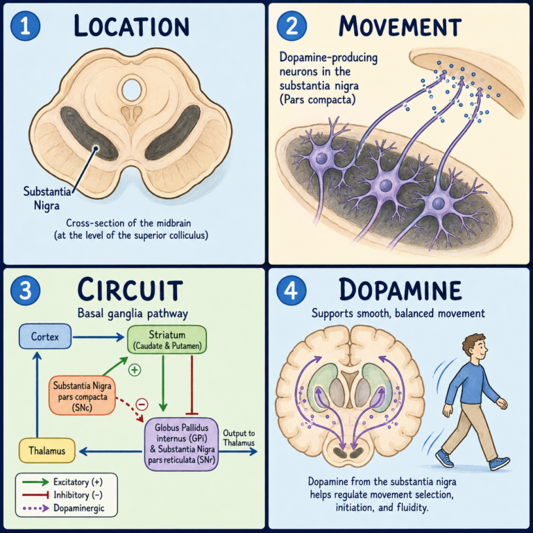
Many diseases affect autonomic function, including diabetic autonomic neuropathy, multiple system atrophy, pure autonomic failure, Parkinson’s disease with autonomic dysfunction, and orthostatic hypotension. People may develop dizziness, abnormal sweating, constipation, urinary problems, abnormal heart-rate control, or impaired blood pressure regulation.
This article is educational background, not a diagnostic guide. Fainting, persistent dizziness on standing, or other ongoing symptoms like these should be evaluated by a clinician.
Related: What Is the Peripheral Nervous System · What Is the Somatic Nervous System? · What Is the Sympathetic Nervous System? · What Is the Parasympathetic Nervous System?
References
- Anatomy, Autonomic Nervous System. StatPearls. NCBI Bookshelf.
- Standring S, ed. Gray’s Anatomy: The Anatomical Basis of Clinical Practice. 42nd ed. Elsevier; 2020.
- Caire MJ, Reddy V, Varacallo MA. Physiology, Synapse. StatPearls. Updated March 27, 2023.
Written by: MedMaru Editorial Team
Reviewed for medical accuracy by:
S. Chang, KMD