Striatum: Location, Function, and Anatomy
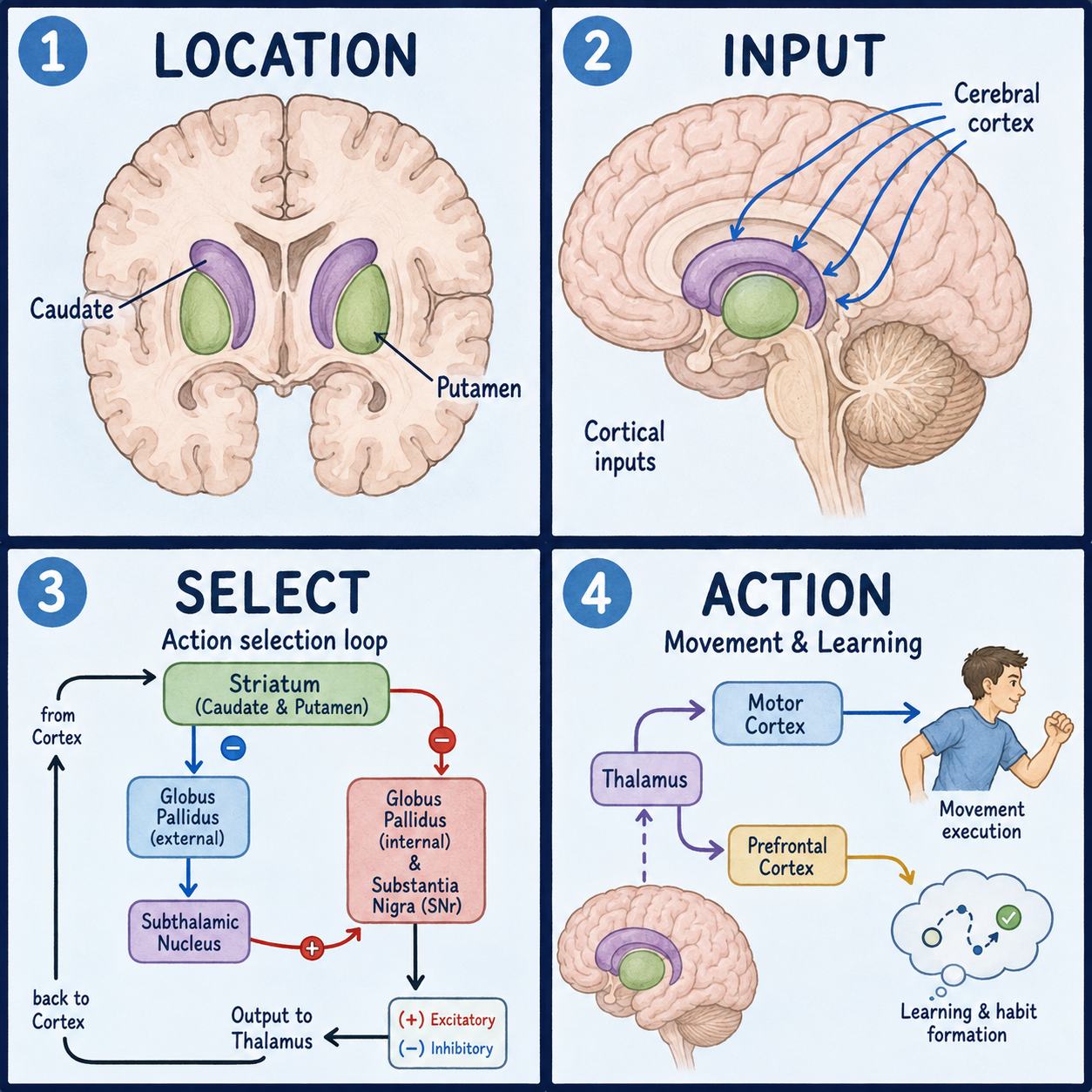
The striatum is a pair of structures buried deep in the brain, one on each side, and it serves as the main entry point for the basal ganglia. The basal ganglia are a group of connected structures beneath the cerebral cortex that shape movement, habits, and motivation. The striatum is built from two parts: the caudate nucleus, a curved, tail-like structure, and the putamen, a rounded structure beside it. Most descriptions also include a lower region called the ventral striatum, which is home to the nucleus accumbens.
If the name sounds familiar, it is probably because of disease. Damage to the striatum, or to the dopamine signal that reaches it, sits at the center of both Parkinson’s disease and Huntington’s disease. Neither condition, though, is explained by this one structure alone.
Why two pieces are really one
On a brain scan, the caudate and putamen look like two separate pieces. They aren’t. They begin as one continuous sheet of tissue, and they only appear split because a band of white matter called the internal capsule grows through the middle of it as the brain develops. Picture a U-shaped gutter pinched in the middle by a cable running through it. The two halves still connect above and below that cable. That is why anatomists treat the caudate and putamen as one working unit, split in appearance but not in function. Like most analogies, this one is only about shape; it says nothing about how the tissue works.
Anatomists do divide the striatum, but by what each zone is for rather than where it sits. The dorsal striatum, made of the caudate and putamen, is tied to conscious movement control and habit formation. The ventral striatum, made of the nucleus accumbens and the olfactory tubercle, is tied to reward, motivation, and emotional processing. There is no sharp wall between the two. Information moves freely across the boundary, and that is part of the reason dopamine problems can affect movement, mood, and motivation at the same time.
Finding it inside the brain
The striatum lies deep inside each cerebral hemisphere. It sits medial to the insula, meaning closer to the midline, and lateral to the thalamus, meaning farther from it. The caudate is shaped like the letter C and curves around the thalamus. Its head bulges into the lateral ventricle, one of the brain’s fluid-filled spaces. Its body runs along the top of the thalamus, and its tail arcs down into the temporal lobe near the amygdala.
The putamen sits just outside the head of the caudate. It pairs with a neighbor, the globus pallidus, inside a lens-shaped unit called the lentiform nucleus. Because the caudate follows the same curve as the lateral ventricle, radiologists often use it as a landmark when they read brain scans.
A station with many lines in and two lines out
The striatum works a little like a train station where several lines arrive but only two depart.
Most of the incoming traffic comes from the cerebral cortex. Nearly the whole cortex sends activating signals into the striatum. Frontal and parietal areas, which handle planning and eye movement, feed mainly into the caudate. Motor and sensory areas, which handle carrying out movement, feed mainly into the putamen. A second line comes from dopamine-producing cells in the substantia nigra. Their fibers form the nigrostriatal pathway, and they fine-tune how the striatum responds to the signals arriving from the cortex. This is the pathway that fails in Parkinson’s disease. The thalamus adds a third input.
The outgoing traffic is carried by the striatum’s main neurons, called medium spiny neurons. They send inhibitory signals onward to the two segments of the globus pallidus, forming what are known as the direct and indirect pathways for movement control. When one incoming line loses its signal, as the dopamine line does in Parkinson’s, the station doesn’t shut down. It starts routing traffic unevenly, and movement becomes harder to start and to control smoothly. The train-station picture has a limit worth stating: real circuits don’t run on fixed schedules, and the balance between pathways shifts continuously.
What goes wrong when it is damaged
In Parkinson’s disease, dopamine-producing neurons in the substantia nigra die off, and the putamen in particular is starved of dopamine input. That tips the balance between the direct and indirect pathways toward less movement and more inhibition, which contributes to slowness and rigidity. Parkinson’s involves much more than the striatum, so describing it as a single-structure disease is never accurate. It is also a common mistake to think dopamine loss only affects movement. Dopamine reaches both the dorsal striatum, which is tied to movement, and the ventral striatum, which is tied to reward, so dopamine-related conditions can affect motivation and mood as well as motor control.
Huntington’s disease works differently. Neurons in the caudate and putamen themselves progressively die, and the loss hits the indirect pathway harder than the direct one. That imbalance is thought to underlie chorea, the involuntary, dance-like movements typical of the disease. Shrinking of the caudate on imaging tracks with declining executive function, but this is a general association from the clinical literature. It is not a rule for predicting any one person’s symptoms from a scan.
This is also why damage to the striatum does not always look the same. The effect depends on which part is affected and which connecting pathway is disrupted. Parkinson’s and Huntington’s both involve the striatum, yet they produce very different, sometimes opposite, movement problems.
The putamen matters in a third, more abrupt way. It is the most common site of hypertensive brain hemorrhage, because of the small perforating arteries that feed it. That is anatomical context, not a diagnostic rule. Beyond these conditions, dysfunction of the caudate and putamen has been linked in the literature to dementia, OCD, ADHD, bipolar disorder, and schizophrenia. These are associations still under study, not established causes, and they are not a self-diagnosis checklist.
One last point of vocabulary helps avoid confusion. The striatum and the basal ganglia are not the same thing. The striatum is one part of the basal ganglia, which also include the globus pallidus, the substantia nigra, and the subthalamic nucleus. The striatum is simply the part that receives most of the incoming signals before the rest of the circuit processes them.
This article is educational background, not a diagnostic guide. Sudden weakness, a severe headache, or a sudden loss of coordination can signal a stroke or hemorrhage and needs urgent medical attention. Tremor, stiffness, slowness, or unexplained involuntary movements should be evaluated by a clinician rather than matched against this article.
Related: What Is the Cerebral Cortex? · What Is the Brain? · What Is the Cerebrum?
References
- StatPearls, “Neuroanatomy, Caudate Nucleus” — https://www.ncbi.nlm.nih.gov/books/NBK557407/
- StatPearls, “Neuroanatomy, Basal Ganglia” — https://www.ncbi.nlm.nih.gov/books/NBK537141/
- Salgado S, Kaplitt MG, “Neuroanatomy of Reward: A View from the Ventral Striatum,” NCBI Bookshelf — https://www.ncbi.nlm.nih.gov/books/NBK92777/
- StatPearls, “Neuroanatomy, Putamen” — https://www.ncbi.nlm.nih.gov/books/NBK542170/
- Purves et al., “Projections to the Basal Ganglia,” Neuroscience, NCBI Bookshelf — https://www.ncbi.nlm.nih.gov/books/NBK10988/
Written by: MedMaru Editorial Team
Reviewed for medical accuracy by:
S. Chang, KMD