What Is a Motor Neuron? Structure, Function, and Types
What Is a Motor Neuron?
Introduction
Every voluntary movement—from taking a step to smiling or lifting a cup—depends on motor neurons.
Motor neurons carry commands from the central nervous system to muscles, allowing the body to move. They form the final pathway through which the brain and spinal cord control skeletal muscles.
Without motor neurons, the brain could generate movement plans, but those commands would never reach the muscles.
Understanding motor neurons is essential for learning how movement is produced and why neurological diseases such as amyotrophic lateral sclerosis (ALS) can cause progressive muscle weakness.
A motor neuron, also called an efferent neuron, is a nerve cell that carries signals from the central nervous system to muscles or glands. Somatic motor neurons control skeletal muscles, while autonomic motor neurons regulate smooth muscle, cardiac muscle, and glands.
What Is a Motor Neuron?
Motor neurons are one of the three major functional classes of neurons.
They form the efferent pathway, carrying information away from the central nervous system to target tissues.
Motor neurons transform decisions made by the brain and spinal cord into muscle contraction or gland secretion.
Types of Motor Neurons
Motor neurons can be classified into two major categories.
Somatic Motor Neurons
Somatic motor neurons innervate skeletal muscles and control voluntary movement.
These neurons produce actions such as:
- Walking
- Writing
- Speaking
- Smiling
- Grasping objects
Each lower motor neuron forms a neuromuscular junction with skeletal muscle fibers.
Autonomic Motor Neurons
Autonomic motor pathways regulate:
- Smooth muscle
- Cardiac muscle
- Glands
Unlike somatic motor neurons, autonomic pathways use two neurons:
- Preganglionic neuron
- Postganglionic neuron
These pathways regulate involuntary functions such as heart rate, digestion, and pupil size.
Upper and Lower Motor Neurons
Motor control depends on two levels of neurons.
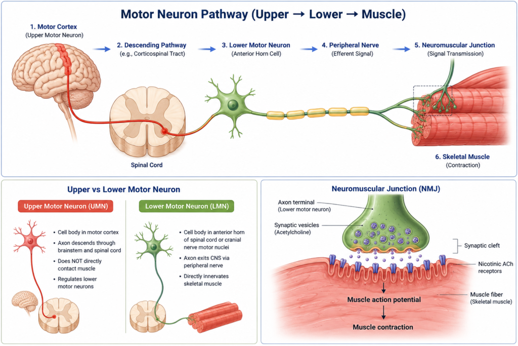
Upper Motor Neurons (UMNs)
Upper motor neurons originate primarily in the primary motor cortex.
Their axons descend through pathways such as the corticospinal tract to reach the brainstem or spinal cord.
Upper motor neurons do not directly contact skeletal muscles.
Instead, they regulate lower motor neurons.
Lower Motor Neurons (LMNs)
Lower motor neurons are located in:
- The anterior horn of the spinal cord
- Motor nuclei of cranial nerves in the brainstem
Their axons leave the central nervous system through peripheral nerves and directly innervate skeletal muscle fibers.
The lower motor neuron is the final common pathway for voluntary movement.
How Motor Neurons Produce Movement
Motor control generally follows these steps.
Step 1. Motor Planning
The brain plans a movement.
↓
Step 2. Upper Motor Neuron Activation
Upper motor neurons transmit descending commands through the brainstem and spinal cord.
↓
Step 3. Lower Motor Neuron Activation
Lower motor neurons receive these signals and generate action potentials.
↓
Step 4. Neuromuscular Junction
Acetylcholine is released at the neuromuscular junction.
↓
Step 5. Muscle Contraction
The muscle fiber depolarizes, initiating contraction.
Neuromuscular Junction
The neuromuscular junction is a specialized synapse between a lower motor neuron and a skeletal muscle fiber.
When an action potential reaches the axon terminal:
- Acetylcholine is released.
- Nicotinic acetylcholine receptors are activated.
- A muscle action potential is generated.
- Muscle contraction begins.
Each lower motor neuron may control from a few to hundreds of muscle fibers, depending on the precision required.
Why Motor Neurons Are Important
Motor neurons allow the brain to convert intention into action.
Without functioning motor neurons, voluntary movement would not be possible.
They are essential for:
- Walking
- Speaking
- Breathing
- Swallowing
- Facial expression
- Fine motor control
Even automatic movements such as posture require continuous motor neuron activity.
Clinical Relevance
Motor neuron disorders can cause weakness, muscle wasting, or paralysis.
Examples include:
- Amyotrophic lateral sclerosis (ALS)
- Poliomyelitis
- Spinal muscular atrophy (SMA)
- Peripheral nerve injury
Upper and lower motor neuron lesions produce different neurological signs, helping clinicians identify the location of injury.
Common Misunderstandings
“Motor neurons only exist in the spinal cord.”
Not true.
Lower motor neurons are found in the spinal cord and in motor nuclei of the brainstem.
“Upper motor neurons directly stimulate muscles.”
Incorrect.
Upper motor neurons control lower motor neurons, which directly innervate skeletal muscles.
“Motor neurons only control voluntary movement.”
Not entirely.
Somatic motor neurons control voluntary skeletal muscles, while autonomic motor pathways regulate involuntary targets such as smooth muscle, cardiac muscle, and glands.
- Motor neurons carry signals from the central nervous system to muscles or glands.
- They are also called efferent neurons.
- Somatic motor neurons directly innervate skeletal muscles.
- Voluntary movement depends on both upper and lower motor neurons.
- The neuromuscular junction converts neural signals into muscle contraction.
Related Articles
- What Is a Sensory Neuron?
- What Is an Interneuron?
- What Is the Somatic Nervous System?
- What Is a Reflex Arc?
References
- Neuroanatomy, Motor Neuron. StatPearls. NCBI Bookshelf.
- Akinrodoye MA, Lui F. Neuroanatomy, Somatic Nervous System. StatPearls. Updated November 7, 2022.
- Grider MH, Jessu R, Kabir R. Physiology, Action Potential. StatPearls. Updated May 8, 2023.
Written by: MedMaru Editorial Team
Reviewed for medical accuracy by:
S. Chang, KMD