Pons: Location, Function, and Anatomy
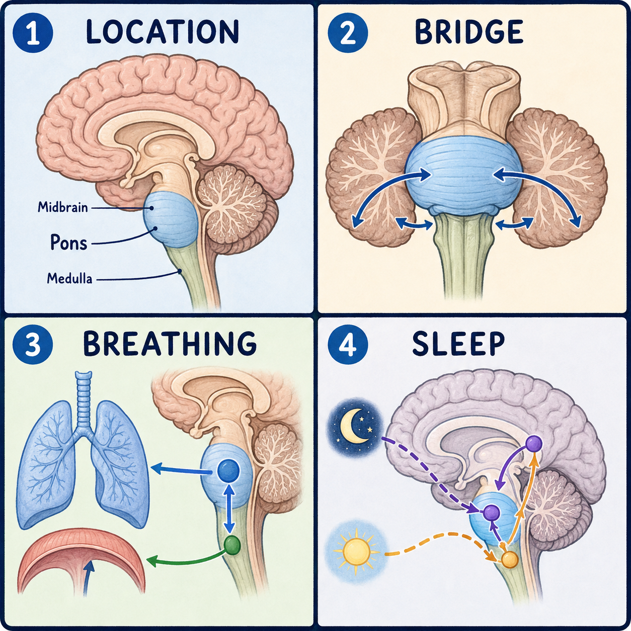
The pons is the middle section of the brainstem, sitting between the midbrain above and the medulla oblongata below. Its name is Latin for “bridge,” and that is a good description of its job. It links the two halves of the cerebellum and carries signals between the brain and the rest of the nervous system. Four cranial nerves connect here, so damage to the pons often produces a particular combination of facial, eye, and hearing problems along with weakness of the body.
Why it is called a bridge
The pons is marked off by two grooves: the pontomesencephalic sulcus at its upper border with the midbrain and the pontomedullary sulcus at its lower border with the medulla. Its most visible feature is a thick bundle of fibers on its front surface that curves around like an arch, connecting to the two sides of the cerebellum through structures called the middle cerebellar peduncles.
It works like a busy highway interchange. Fibers from the cerebral cortex pass straight through on their way down to the spinal cord, while other traffic turns off sideways into the cerebellum. The interchange image fits the layout, though unlike a road junction, the pons also contains its own control centers, as described below.
Where it sits, and why that matters
The pons lies in front of the fourth ventricle and behind the clivus, a sloping part of the skull base. The midbrain is above it, the medulla below, and the cerebellum just behind.
This crowded location has a serious clinical side. The basilar artery runs along the front of the pons and supplies it with blood. If that artery becomes blocked, the resulting stroke can damage a wide area of the pons and, in severe cases, cause locked-in syndrome. A person with locked-in syndrome is conscious and aware but almost completely unable to move or speak. This happens because damage to the front, or ventral, pons cuts the descending motor pathways, the corticospinal and corticobulbar tracts, while sparing the reticular activating system toward the back that keeps a person conscious. Vertical eye movements, which are controlled from the midbrain above the damage, are often preserved, while horizontal eye movements, which depend on the abducens nucleus within the affected pons, are typically lost. That pattern helps clinicians place the problem in the pons, and it matters because someone who looks unresponsive may be fully aware.
Front and back: two different jobs
In cross-section, the pons has two regions. The basilar, or ventral, pons faces forward and mainly acts as the bridge. It contains the pontine nuclei, which receive signals from the cerebral cortex and pass them sideways into the cerebellum, and it carries corticospinal fibers, movement commands bound for the spinal cord, that simply pass through.
The pontine tegmentum sits toward the back. Like the tegmentum of the midbrain, it houses most of the pons’s cranial nerve nuclei along with part of the reticular formation, a network of neurons that helps regulate arousal. Working with centers in the medulla, this network modulates the strength and rhythm of breathing rather than generating the rhythm itself. So it is a misconception to think the pons only relays traffic to the cerebellum; it also runs control centers of its own.
One landmark in the tegmentum is the facial colliculus, a small bump on the floor of the fourth ventricle. It isn’t the facial nerve nucleus itself. It is formed by facial nerve fibers looping around the abducens nucleus underneath, a bit like a cable wrapped around a pipe. That detail explains why some pontine injuries damage facial movement and eye movement together, even though the two nerves leave the brainstem separately.
Four cranial nerves
Four of the twelve cranial nerves have nuclei in the pons. The trigeminal nerve, cranial nerve V, carries most facial sensation and controls the chewing muscles, leaving the pons as a larger sensory root and a smaller motor root. The abducens nerve, VI, controls the lateral rectus muscle, which turns the eye outward. The facial nerve, VII, controls the muscles of facial expression. The vestibulocochlear nerve, VIII, carries hearing and balance information, and its nuclei extend across the border between the pons and the medulla.
What damage looks like
Because the pons is a narrow crossroads for movement, sensation, and cranial nerve pathways, the effects of damage depend heavily on where it falls. Damage toward the front mainly disrupts the descending motor fibers and causes weakness. Damage toward the back, near the cranial nerve nuclei, is more likely to affect eye movement, facial sensation or movement, or hearing and balance, and it can involve nearby cerebellar pathways, causing poor coordination.
Outcomes vary just as widely. Some pontine strokes cause limited symptoms that improve, while a large stroke across the ventral pons can cause locked-in syndrome or be life-threatening, since the pons also contributes to basic functions such as breathing regulation. That is general information, not a prediction for any individual.
This article is educational background, not a diagnostic guide. Sudden weakness, double vision, facial drooping, slurred speech, or severe dizziness needs emergency medical care.
Related: What Is the Cerebral Cortex? · What Is the Brain? · What Is the Brainstem?
References
- StatPearls. “Neuroanatomy, Pons.” NCBI Bookshelf, NBK560589 — https://www.ncbi.nlm.nih.gov/books/NBK560589/
- StatPearls. “Neuroanatomy, Locked-In Syndrome.” NCBI Bookshelf, NBK559026 — https://www.ncbi.nlm.nih.gov/books/NBK559026/
- StatPearls. “Physiology, Pontine.” NCBI Bookshelf, NBK482414 — https://www.ncbi.nlm.nih.gov/books/NBK482414/
Written by: MedMaru Editorial Team
Reviewed for medical accuracy by:
S. Chang, KMD