Medulla Oblongata: Location, Function, and Anatomy
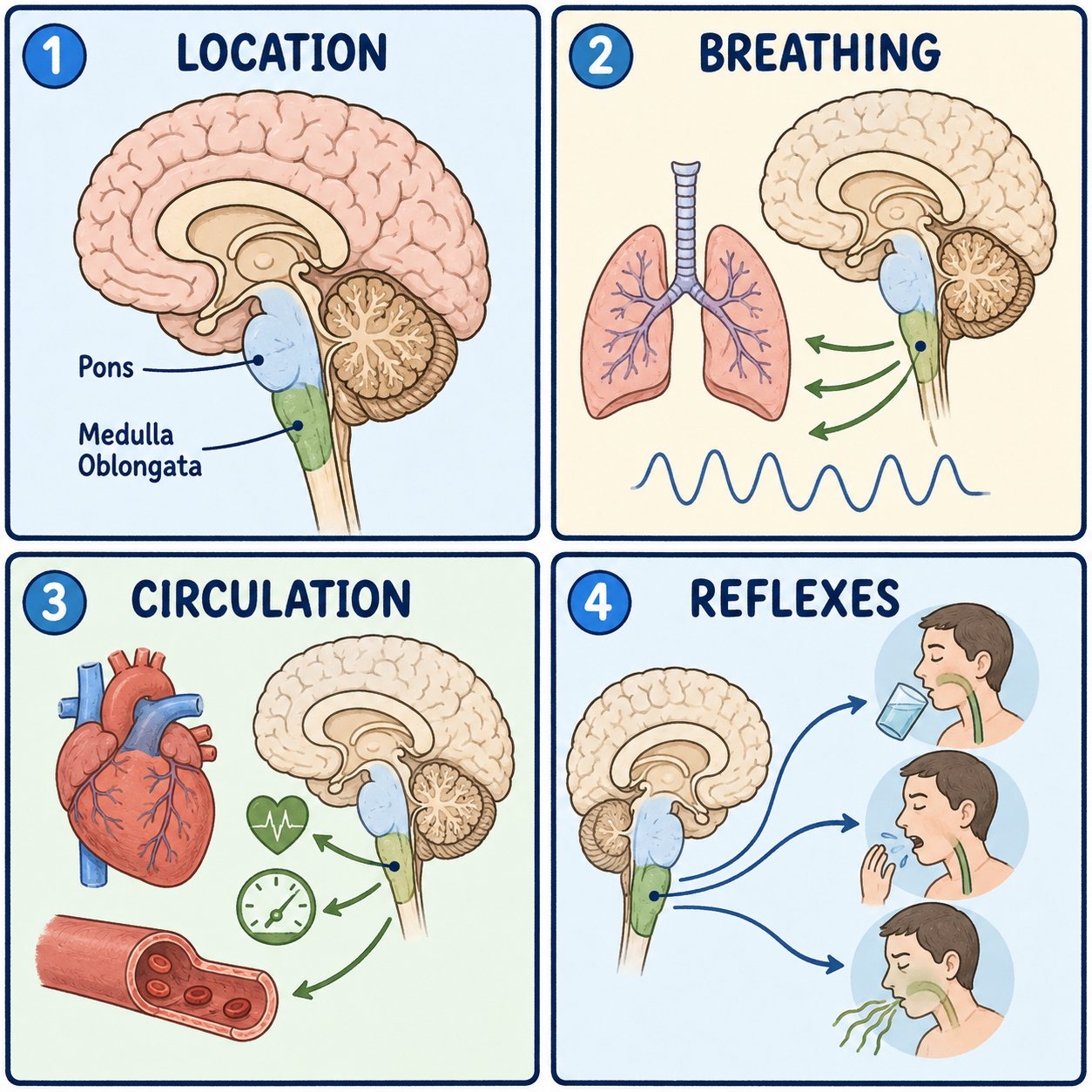
The medulla oblongata is the lowest section of the brainstem, the point where the brain becomes the spinal cord. It works like a life-support control room. It houses the centers that keep breathing rhythm and heart rate running automatically, without any conscious effort, and it is also where most movement signals from one side of the brain cross over to control the opposite side of the body. Because it manages such basic survival functions, damage here is taken very seriously in medicine.
A thickened continuation of the spinal cord
The medulla, often just called “the medulla,” is the lowest of the brainstem’s three parts. It sits roughly at the level of the foramen magnum, the large opening in the base of the skull where the brain connects to the spinal cord. On its front surface, a groove called the anterior median fissure lines up directly with the same groove running down the spinal cord. It is a visible reminder that the medulla is essentially the spinal cord continued upward, thickened and reorganized to hold extra control centers. Its lower part looks much like the cord itself, while its upper part opens out to form the floor of the fourth ventricle, which it shares with the pons above. So it is part of the brainstem, not the whole of it; the midbrain and pons sit above.
Little room to spare
The medulla sits below the pons and above the spinal cord, with the boundary usually placed at the foramen magnum. Being surrounded so closely by bone is protective, but it also leaves very little room for swelling. That matters because the medulla lies at the very bottom of the skull, right where rising pressure inside the head tends to push tissue downward. Severe brain swelling can squeeze the medulla against the foramen magnum, which is life-threatening because it can shut down the centers that keep a person breathing.
The landmarks that do the work
A handful of landmarks account for most of what the medulla does. On its front surface, two raised ridges called the pyramids carry the corticospinal tract, the main pathway for voluntary movement commands from the brain to the spinal cord. Near the bottom of the medulla, most of these fibers cross to the other side at the pyramidal decussation, a bit like a highway interchange where most traffic switches to the opposite carriageway. This crossing is the main reason a stroke on one side of the brain usually causes weakness on the opposite side of the body.
Toward the back, the gracile and cuneate nuclei receive information about touch, vibration, and body position coming up from the spinal cord and relay it through a pathway called the medial lemniscus toward the thalamus and on to the cortex. The nucleus of the solitary tract receives and coordinates sensory information from the internal organs, including blood pressure, stomach fullness, and blood oxygen levels, which makes it a key hub for the body’s automatic functions. Nearby groups of neurons, sometimes described together as the ventral respiratory column and the rostral ventrolateral medulla, set the basic rhythm of breathing and help maintain baseline blood pressure, adjusting moment to moment in response to signals such as oxygen and carbon dioxide levels in the blood. (For how these stations work together, see how the brain controls the autonomic nervous system.)
The area postrema, a small structure toward the back, sits outside the usual blood-brain barrier, so it can directly sense toxins and chemicals circulating in the blood. Acting as a chemoreceptor trigger zone rich in dopamine receptors, it plays a central role in triggering vomiting when something harmful is detected in the bloodstream. It is also a target of certain anti-nausea medicines, including some used for chemotherapy-induced nausea, which involves serotonin, substance P, and dopamine receptors in this region.
All of this is why the medulla is far more than a cable between the brain and spinal cord. It is structurally continuous with the cord, but it houses dedicated control centers for breathing, blood pressure, swallowing, and vomiting.
The cranial nerves that connect here
Four of the twelve cranial nerves connect to the medulla. The glossopharyngeal nerve, IX, helps control swallowing and carries some taste and throat sensation. The vagus nerve, X, sends parasympathetic signals to the heart, lungs, stomach, and much of the digestive tract, and it contributes to the gag and cough reflexes. The accessory nerve, XI, controls two neck and shoulder muscles. The hypoglossal nerve, XII, controls the muscles of the tongue, and damage to it weakens the tongue on the same side.
When the medulla is damaged
Because motor, sensory, and autonomic pathways are packed so tightly here, damage can range from limited and manageable to immediately life-threatening, depending on exactly which structures are involved.
Lateral medullary syndrome, also called Wallenberg syndrome, is the most common type of medullary stroke and usually results from a blockage in the vertebral artery. It affects the outer part of the medulla rather than the pyramids, so it typically does not cause limb weakness. Instead it causes difficulty swallowing, vertigo, loss of pain and temperature sensation on one side of the face and the opposite side of the body, and a set of eye and eyelid changes on the affected side known as Horner syndrome. It is a useful reminder that not every stroke causes weakness on one side; a stroke can badly affect sensation, swallowing, and coordination while sparing limb strength, because those pathways are separate from the corticospinal tract.
Medial medullary syndrome, or Dejerine syndrome, results from damage nearer the midline. It typically causes weakness of the arm and leg on the opposite side, because the pyramid is damaged above the point where its fibers cross, together with tongue weakness on the same side, because the hypoglossal nerve fibers on their way out of the brainstem are damaged directly.
Because of the medulla’s role in survival functions, injuries or surgery near it call for careful monitoring of breathing and heart rhythm afterward.
This article is educational background, not a diagnostic guide. Sudden difficulty swallowing, severe vertigo, new numbness or weakness, or trouble breathing needs emergency medical care.
Related: What Is the Cerebral Cortex? · What Is the Brain? · What Is the Brainstem?
References
- StatPearls. “Neuroanatomy, Medulla Oblongata.” NCBI Bookshelf, NBK551589 — https://www.ncbi.nlm.nih.gov/books/NBK551589/
- StatPearls. “Physiology, Chemoreceptors” (Area Postrema). NCBI Bookshelf, NBK544249 — https://www.ncbi.nlm.nih.gov/books/NBK544249/
- National Cancer Institute PDQ. “Nausea and Vomiting.” — https://www.cancer.gov/about-cancer/treatment/side-effects/nausea/nausea-hp-pdq
Written by: MedMaru Editorial Team
Reviewed for medical accuracy by:
S. Chang, KMD