Midbrain: Location, Function, and Anatomy
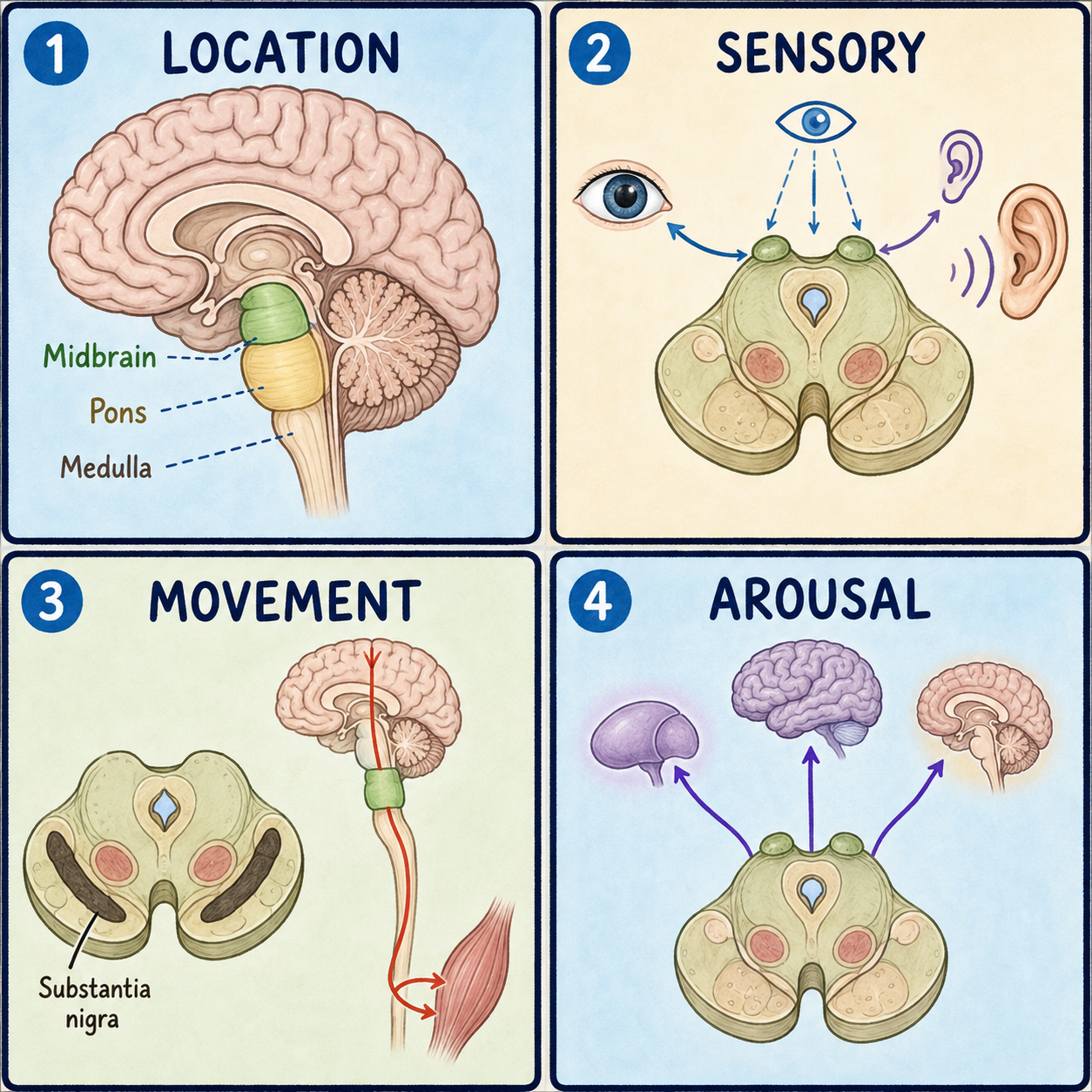
The midbrain, also called the mesencephalon, Greek for “middle brain,” is the smallest of the brainstem‘s three parts. It sits between the upper brain and the lower brainstem and works like a relay station and control tower in one. Visual and auditory signals pass through it, eye movements are coordinated there, and a cluster of dopamine-producing cells called the substantia nigra helps make smooth, voluntary movement possible. Because so many pathways converge at this narrow point, even a small midbrain injury tends to produce a distinctive combination of symptoms rather than one isolated problem.
A narrow crossroads at the top of the brainstem
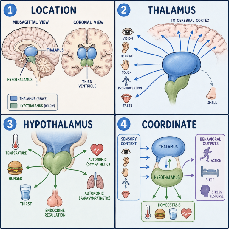
The midbrain is roughly 1.5 centimeters long and forms the top section of the brainstem, which connects the cerebrum to the spinal cord in three stacked parts: midbrain on top, pons in the middle, and medulla oblongata at the bottom. So the midbrain is part of the brainstem, not the whole of it. It sits above the pons and below the diencephalon, the region containing the thalamus and hypothalamus, and its lower border meets the pons at a visible groove called the pontomesencephalic sulcus. On each side it lies close to the temporal lobes, and it passes through an opening in the tentorium cerebelli, a tough sheet of tissue separating the cerebrum from the cerebellum.
A thin canal called the cerebral aqueduct runs through the center of the midbrain, carrying cerebrospinal fluid from the third ventricle above to the fourth ventricle below. Because this channel is so narrow, it is one of the places where fluid flow can most easily become blocked, which is one cause of hydrocephalus, a buildup of fluid in the brain.
The location has another clinical consequence. When pressure rises inside the skull, from bleeding or a large mass for example, brain tissue can be pushed downward through the tentorial opening and squeeze the midbrain. This is called uncal or transtentorial herniation. The oculomotor nerve running through this area carries the parasympathetic fibers that keep the pupil constricted, and these fibers tend to be compressed first. That leaves the pupil-widening sympathetic fibers unopposed, which is why one of the earliest warning signs of herniation is a pupil on one side becoming wide and unresponsive to light.
The roof and the floor: tectum and tegmentum
In cross-section, the midbrain divides into two main regions. The tectum, Latin for “roof,” forms the back part. It carries four small bumps, the corpora quadrigemina, arranged in two pairs. The superior colliculi help direct eye and head movements toward things you see, and the inferior colliculi relay sound information toward the hearing areas of the cortex. You can think of the tectum as a traffic controller for “look here” and “listen to that” reflexes, though the comparison covers only its reflexive role and leaves out the deliberate processing that happens elsewhere in the brain.
The tegmentum, Latin for “covering,” forms the front and central part, and it houses several structures important for movement and alertness. The red nucleus, which gets its pinkish color from iron-containing pigment, helps coordinate limb movement. The substantia nigra, a dark band of dopamine-producing cells, is part of the basal ganglia movement circuit. It is a normal part of everyone’s movement control and becomes clinically important when its cells degenerate, as in Parkinson’s disease. The periaqueductal gray surrounds the cerebral aqueduct and is one of the brain’s built-in pain-control centers, releasing natural opioid-like chemicals called enkephalins that dampen pain signals. Part of the reticular formation, a network of neurons running through the whole brainstem that helps keep you awake and alert, also passes through here.
In front of the tegmentum lie the cerebral peduncles, also called the crus cerebri. These thick bundles of nerve fibers carry voluntary movement commands from the cerebral cortex down toward the spinal cord, acting as the midbrain’s on-ramp for descending motor signals.
This is why it’s a misconception to treat the midbrain as just a corridor. It actively processes visual and auditory reflexes through the colliculi and pain signals through the periaqueductal gray, rather than merely passing signals between the cerebrum and the body.
Two cranial nerves begin here
Two of the twelve cranial nerves have their control centers, or nuclei, in the midbrain. The oculomotor nerve, cranial nerve III, controls most eye movements and constriction of the pupil. The trochlear nerve, cranial nerve IV, controls a single eye muscle, the superior oblique, and is unusual as the only cranial nerve that exits from the back of the brainstem rather than the front.
What damage looks like
Because so many pathways share such a small space, midbrain injury rarely causes just one problem. Effects depend on the exact location and extent, but they commonly include problems with eye movement or pupil response, weakness on one side of the body, or reduced alertness.
Weber syndrome occurs when a stroke damages the front, inner part of the midbrain, affecting both the oculomotor nerve fibers and the movement pathway in the cerebral peduncle. The result is a drooping eyelid and an outward-turned eye on the same side as the injury, paired with weakness on the opposite side of the body. The pattern is crossed because the motor fibers cross to the other side farther down, in the lower brainstem and spinal cord.
Parinaud syndrome results from pressure on or damage to the tectum, most often from a tumor in the nearby pineal region. Because the superior colliculi help drive upward eye movement, the classic sign is difficulty looking up.
Parkinson’s disease is linked to progressive loss of the dopamine-producing cells in the substantia nigra, typically more than 50 percent and up to about 80 percent by the time motor symptoms such as tremor, stiffness, and slowed movement appear. Parkinson’s involves more than one brain region and more than one neurotransmitter system, though, so describing it simply as a substantia nigra disease oversimplifies a broader process that is still not fully understood.
Not all brainstem strokes are alike, either. Strokes in the midbrain, pons, and medulla produce different symptom patterns depending on which structures are involved, and severity depends on exact location and extent as much as on which of the three regions is affected.
This article is educational background, not a diagnostic guide. A suddenly drooping eyelid, double vision, an unequal pupil, weakness on one side, or reduced alertness needs emergency medical care.
Related: What Is the Cerebral Cortex? · What Is the Brain? · What Is the Brainstem?
References
- StatPearls. “Neuroanatomy, Mesencephalon Midbrain.” NCBI Bookshelf, NBK551509 — https://www.ncbi.nlm.nih.gov/books/NBK551509/
- StatPearls. “Uncal Herniation.” NCBI Bookshelf, NBK537108 — https://www.ncbi.nlm.nih.gov/books/NBK537108/
- StatPearls. “Parkinson Disease.” NCBI Bookshelf, NBK470193 — https://www.ncbi.nlm.nih.gov/books/NBK470193/
Written by: MedMaru Editorial Team
Reviewed for medical accuracy by:
S. Chang, KMD