Thalamus vs. Hypothalamus: What’s the Difference
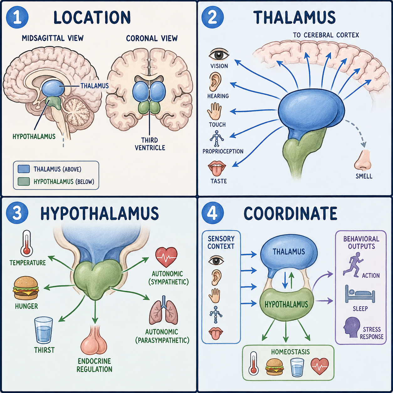
The thalamus and hypothalamus are two small structures near the center of the brain that sit right next to each other but do very different jobs. The thalamus works like a switching hub. It passes almost all sensory information, what you see, hear, and feel, up to the thinking parts of the brain, and it helps control how alert you are. The hypothalamus sits just below it and works more like a control room for the body, keeping internal conditions steady by adjusting body temperature, hunger, thirst, sleep timing, and stress hormones. Both belong to a deep, midline region called the diencephalon. Put simply, the thalamus mainly manages what information reaches awareness, while the hypothalamus mainly manages the body’s automatic, moment-to-moment survival needs.
Neighbors with different jobs
The thalamus is a paired, egg-shaped mass of gray matter, tissue made mostly of nerve cell bodies, lying just above the midbrain. There is one on each side of the brain, and together they form much of the side walls of a fluid-filled space called the third ventricle.
The hypothalamus is smaller. Its name literally means “below the thalamus,” and it surrounds the lower part of the third ventricle. Its boundaries are a thin membrane at the front called the lamina terminalis, the pituitary gland below, the mammillary bodies at the back, and a shallow groove called the hypothalamic sulcus that separates it from the thalamus above.
A useful picture: the thalamus is like an airport control tower deciding which incoming flights, the sensory signals, are cleared to land at their gates in the cortex. The hypothalamus is like the facilities office quietly running the heating, water, and security systems. The analogy has a limit. A control tower and a facilities office don’t share a wall or exchange direct signals, while the thalamus and hypothalamus are physically adjacent and directly linked by fiber tracts, including the mammillothalamic tract.
The thalamus: more than a relay station
The thalamus is bordered on its outer side by the internal capsule, a major band of white matter, with the hypothalamus below. Internal sheets of white matter called the medullary laminae divide it into anterior, medial, and lateral groups of nuclei, clusters of neurons that share a function.
Its named nuclei show that the thalamus is really a set of specialized relay stations. The lateral geniculate nucleus relays visual signals from the retina toward the visual cortex in the occipital lobe. The medial geniculate nucleus relays sound signals from the inferior colliculus toward the hearing cortex in the temporal lobe. The ventral posterior nucleus relays body sensation, including pain, temperature, and touch arriving through the spinothalamic tract, as well as facial sensation from the trigeminal nerve. Because nearly every sense passes through the thalamus on its way to the cortex, smell being the exception, a small thalamic lesion can cause a surprisingly large and precise loss of sensation on the opposite side of the body.
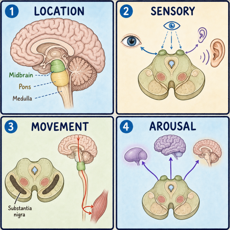
Calling the thalamus a passive relay would still be an oversimplification. The reticular nucleus wraps around the rest of the thalamus and actively regulates the other nuclei, helping control alertness and attention. The intralaminar nuclei, which receive input from the brainstem’s reticular activating system, contribute to alertness and consciousness. The thalamus also connects with memory-related structures such as the hippocampus, mammillary bodies, and fornix, and it takes part in sleep regulation. According to NINDS, during most sleep stages the thalamus becomes quiet, letting you tune out the outside world, but during REM sleep it becomes active again and sends the cortex the images, sounds, and other sensations that fill dreams.
The hypothalamus: a thermostat with many dials
The hypothalamus is organized into three zones running outward from the wall of the third ventricle, the periventricular, medial, and lateral zones, which together contain about eleven distinct nuclei. Each handles different regulatory work.
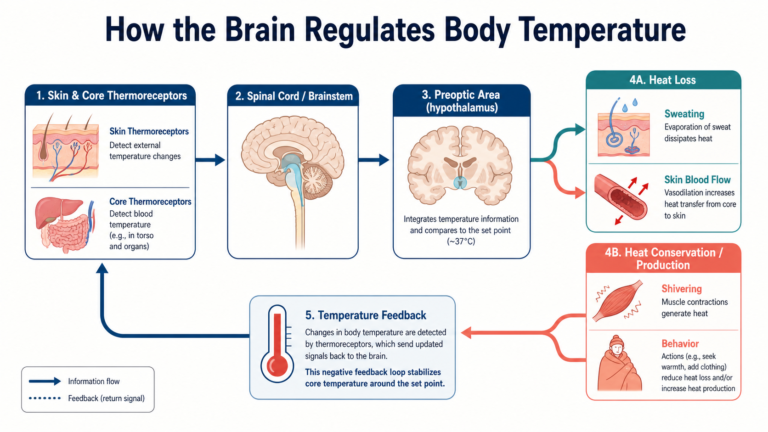
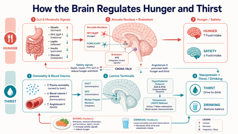
The suprachiasmatic nucleus receives light signals directly from the eyes and sets the roughly 24-hour circadian rhythm. The paraventricular and supraoptic nuclei produce oxytocin and antidiuretic hormone, which are released from the back part of the pituitary gland, and the paraventricular nucleus also releases corticotropin-releasing hormone, which drives the stress-hormone response. The preoptic, anterior, and posterior nuclei regulate body temperature. In animal studies, destroying the ventromedial nucleus produces overeating, linking it to fullness, while damage to the lateral hypothalamus is associated with reduced eating. The arcuate nucleus helps control hormone release from the front part of the pituitary through a dedicated blood-vessel connection, the hypothalamic-pituitary portal system, and it responds to the appetite hormones leptin and ghrelin. The mammillary nuclei take part in a memory loop, the Papez circuit.
A thermostat is a fair comparison, since the hypothalamus senses conditions and switches other systems on or off. Unlike a thermostat, though, it juggles temperature, water balance, hunger, stress hormones, and daily timing all at once. It also isn’t just “the hormone part of the brain.” It controls temperature and autonomic functions through direct nerve pathways to the brainstem and spinal cord, not only through hormones.
Why one nucleus rarely means one symptom
Lists that pair each nucleus with one job are useful for learning, but most of those pairings come from lesion and stimulation studies. In people, damage usually affects several neighboring nuclei at once, so it tends to produce overlapping symptoms rather than one clean deficit. A lesion reaching both the thalamus and hypothalamus can produce a mix of sensory and regulatory problems.
When these structures are damaged
A thalamic stroke can cause Dejerine-Roussy syndrome, in which initial numbness on one side can, months later, turn into severe chronic pain, a documented pattern that is not fully explained. Thalamic aphasia has its own pattern of word-finding and meaning errors with preserved repetition, and recovery is often relatively fast compared with some aphasias from cortical damage. Thalamic damage can also affect consciousness and sleep. In treatment, deep brain stimulation of a thalamic nucleus called the ventral intermediate nucleus is used for medication-resistant essential tremor and Parkinson’s tremor.
Around the hypothalamus, pituitary adenomas are the most common mass lesions. Because the hypothalamus sits directly above the pituitary and communicates with it both through hormones and through nerve fibers, disease here can disrupt the whole chain. Adenomas can cause hormone excess or deficiency, and they can press on the nearby optic chiasm to cause bitemporal hemianopsia, loss of the outer half of the visual field in both eyes. Disrupted antidiuretic hormone signaling can lead to diabetes insipidus or SIADH, a condition of excess water retention. Wernicke-Korsakoff syndrome, caused by thiamine deficiency, characteristically damages the mammillary bodies.
Whether hypothalamic damage can be reversed depends entirely on the cause, which nuclei are involved, and how quickly treatment starts. Some hormonal deficits can be managed well, while some loss of function can be permanent.
This article describes structure-function relationships from anatomy and physiology references. It is not a diagnostic guide, and conditions in this region need specialist evaluation and imaging.
Related: What Is the Diencephalon? · What Is the Cerebral Cortex? · What Is the Brain?
References
- StatPearls. “Neuroanatomy, Thalamus.” NCBI Bookshelf, NBK542184 — https://www.ncbi.nlm.nih.gov/books/NBK542184/
- StatPearls. “Neuroanatomy, Thalamic Nuclei.” NCBI Bookshelf, NBK549908 — https://www.ncbi.nlm.nih.gov/books/NBK549908/
- StatPearls. “Neuroanatomy, Hypothalamus.” NCBI Bookshelf, NBK525993 — https://www.ncbi.nlm.nih.gov/books/NBK525993/
- NINDS. “Brain Basics: Understanding Sleep.” — https://www.ninds.nih.gov/health-information/public-education/brain-basics/brain-basics-understanding-sleep
Written by: MedMaru Editorial Team
Reviewed for medical accuracy by:
S. Chang, KMD