How Do Neurons Combine Excitatory and Inhibitory Signals?
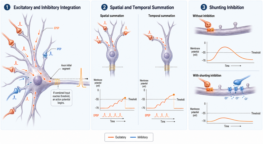
A single neuron can receive thousands of signals from other cells at once. Some push it toward firing, while others hold it back or regulate how much the excitatory signals count. The neuron is constantly combining these inputs before it “decides” whether to send an action potential of its own. This process is called synaptic integration. Inputs arriving at different synapses add up through spatial summation, and inputs arriving close together in time add up through temporal summation. If the combined effect depolarizes a trigger zone called the axon initial segment enough, the neuron fires.
A vote that isn’t a simple count
Most chemical synapses have one of two broad effects. An excitatory postsynaptic potential, or EPSP, makes the neuron more likely to fire. An inhibitory postsynaptic potential, or IPSP, makes it less likely, or limits the effect of excitatory inputs. Both are graded potentials, meaning their size varies, and they usually weaken as they spread through the dendrites and cell body.
It is tempting to imagine the neuron as a ballot box that counts yes and no votes. That is a useful first picture, but real neurons do not simply count active synapses. They weigh each input by its strength, timing, location, receptor type, and effect on the membrane’s conductance. Simply adding excitation and subtracting inhibition leaves much of the story out.
Where the inputs land and where the decision is made
Most synaptic inputs arrive on the dendrites, on small protrusions called dendritic spines, or on the cell body. Some specialized inhibitory synapses target the axon initial segment directly. Dendrites are not passive wires. Their branching shape, ion channels, and electrical properties influence how signals spread and interact. A signal produced far from the cell body may fade as it travels, but distance is not the whole story, because strong synapses and active dendritic channels can amplify or reshape particular inputs.
The final decision to fire is usually made at the axon initial segment, a specialized region near the start of the axon. It is packed with voltage-gated sodium channels, which makes it especially sensitive to depolarization. Simplified explanations often name the axon hillock as the firing site, but in most neurons action potentials start more specifically in the axon initial segment, just beyond the hillock.
How excitation pushes toward threshold
An excitatory synapse produces an EPSP. In many neurons of the central nervous system, the neurotransmitter glutamate activates receptors such as AMPA receptors. The resulting ion flow creates a net inward positive current, which depolarizes the membrane and moves it closer to the threshold for an action potential.
One EPSP on its own is often too small to reach threshold. Several usually need to overlap. Excitation raises the probability of firing, but it does not guarantee it.
Two ways to hold a neuron back
Inhibition reduces the chance of firing or limits the impact of excitation. In the mature central nervous system, many inhibitory synapses release GABA. GABA can activate GABA-A receptors, which are ion channels mainly permeable to chloride, and GABA-B receptors, which act more slowly through G proteins and can influence potassium and calcium channels.
Inhibition works in two important ways. In hyperpolarizing inhibition, the input pushes the membrane potential farther from threshold, so the neuron needs more excitation to fire. In shunting inhibition, the inhibitory signal may cause little visible hyperpolarization. Instead it opens more channels, increasing the membrane’s conductance so that some of the excitatory current leaks away across the membrane. Picture trying to fill a bucket while someone drills extra holes in it: the water still comes in, but less of it stays. The analogy is rough, but it shows why not every inhibitory signal hyperpolarizes the neuron, and why inhibition is not always the subtraction of a fixed amount of voltage. It changes how effectively other signals can influence the cell.
Adding across space and across time
Spatial summation happens when potentials generated at different synapses overlap. Two excitatory neurons might release neurotransmitter onto different parts of the same neuron at about the same moment. Each EPSP may be too small alone, but together they can bring the neuron to threshold. Excitatory and inhibitory inputs interact spatially too. An inhibitory synapse near the cell body or axon initial segment can strongly influence whether excitation leads to firing, while inhibition on one dendritic branch can selectively control the excitatory inputs arriving on that branch.
Temporal summation happens when potentials arrive close together in time. If a new EPSP begins before the previous one has faded, the two overlap, so repeated activation of the same synapse can produce more depolarization than a single input. Repeated IPSPs can overlap in the same way, producing stronger or longer inhibition. How well this works depends partly on how quickly the neuron’s membrane voltage changes and settles back toward rest. In real neurons, spatial and temporal summation usually happen together.
What decides how much an input counts
Several factors shape the effect of any one synaptic signal. Stronger synapses usually produce larger currents, and synaptic strength itself can change through the processes behind learning and plasticity. Timing matters, since signals arriving close together are more likely to overlap, and small timing differences can decide whether the neuron reaches threshold. Location matters, because inputs closer to the cell body or axon initial segment may have a greater effect, though dendritic structure and local channels can change that relationship.
Receptor type matters too. Ionotropic receptors usually act quickly, while metabotropic receptors generally produce slower, longer-lasting effects. The neuron’s existing membrane potential when the signal arrives can change its impact. So can membrane conductance: when more channels are already open, the membrane may respond less to additional inputs, as in shunting inhibition. Finally, dendrites contain voltage-gated channels and can do their own local processing. Under certain conditions, coordinated inputs can trigger local dendritic spikes involving sodium, calcium, or NMDA receptor-dependent currents.
Crossing the threshold
The combined synaptic input shapes the voltage and conductance near the axon initial segment. If the result is enough depolarization to reach threshold, voltage-gated sodium channels open and an action potential starts down the axon. If threshold isn’t reached, no full action potential occurs. Postsynaptic potentials are graded, while action potentials are generally all-or-none. The threshold is not a perfectly fixed voltage, though. It can shift with recent electrical activity, the state of ion channels, and how fast the membrane depolarizes.
This selectivity is what makes integration valuable. Rather than firing whenever any single input is active, neurons can compare competing signals, detect inputs that occur together, filter out irrelevant activity, control the timing of their spikes, process sensory information, select and coordinate movements, adapt with learning, and help keep network activity stable. The interplay between excitation and inhibition lets the nervous system stay responsive without becoming overactive or falling silent.
It also explains why neurotransmitters are not simply excitatory or inhibitory by nature. Glutamate is usually excitatory and GABA usually inhibitory in the mature central nervous system, but the effect depends on the receptor, ion gradients, and cellular context, and these descriptions do not hold in every cell or at every stage of development.
When the balance is disrupted
Disrupted regulation of excitation, inhibition, and neuronal synchronization can contribute to abnormal neural activity, including seizures. Changes in the relationship between excitation and inhibition are also being investigated in several neurological, neurodevelopmental, and psychiatric conditions. There is no single, universal form of excitation–inhibition imbalance, however. The relevant changes can differ between brain regions, neuron and receptor types, circuits, developmental stages, and conditions. Excitation–inhibition imbalance is best understood as a broad research framework, not a complete diagnosis or a single abnormality measured the same way in everyone.
Related: What Are Neurotransmitters? · What Are Excitatory and Inhibitory Signals? · What Are Neural Receptors and How Do They Work? · What Is a Neural Circuit? · What Is Excitation–Inhibition Balance in the Brain? · What Is an Action Potential? Brain Signals Explained
References
- Purves D, Augustine GJ, Fitzpatrick D, et al. Neuroscience. 2nd ed. Sinauer Associates; 2001. Summation of Synaptic Potentials—NCBI Bookshelf
- Purves D, Augustine GJ, Fitzpatrick D, et al. Neuroscience. 2nd ed. Sinauer Associates; 2001. Excitatory and Inhibitory Postsynaptic Potentials—NCBI Bookshelf
- Betts JG, Young KA, Wise JA, et al. Anatomy and Physiology 2e. OpenStax; 2022. Communication Between Neurons
- Huang CYM, Rasband MN. Axon initial segments: structure, function, and disease. Annals of the New York Academy of Sciences. 2018;1420(1):46–61. Full Text—PubMed Central
- Leterrier C. The axon initial segment: an updated viewpoint. Journal of Neuroscience. 2018;38(9):2135–2145. Full Text—PubMed Central
Written by: MedMaru Editorial Team
Reviewed for medical accuracy by:
S. Chang, KMD