Cerebrum: Anatomy, Lobes, and Function
The cerebrum is the largest part of the human brain, the big, folded structure you see when you look at a brain from above or from the side. It is made mainly of the left and right cerebral hemispheres, and each hemisphere has an outer layer called the cerebral cortex, white matter beneath it, and deep clusters of gray matter. The cerebrum lets us sense the world consciously, move on purpose, use language, learn and remember, pay attention, plan and decide, feel emotion and motivation, and recognize objects, faces, and spatial relationships. It doesn’t do any of this alone. These abilities arise from networks spread across the cerebrum and connected with the thalamus, brainstem, cerebellum, spinal cord, and the rest of the nervous system.
The cerebrum is not the whole brain
During embryonic development, the cerebral hemispheres grow from the telencephalon, a division of the developing forebrain. It’s common to use “cerebrum” as another word for the brain, but that’s not accurate. The brain also includes the diencephalon, which holds the thalamus and hypothalamus, as well as the brainstem and the cerebellum. Anatomy texts differ somewhat on where to draw boundaries for deep forebrain structures, but in general each cerebral hemisphere includes the cortex, the underlying white matter, and deep gray-matter structures such as the basal nuclei.
Two hemispheres in constant conversation
A deep groove called the longitudinal fissure separates the left and right hemispheres along most of their upper surface. They are not isolated from each other. Bundles of nerve fibers called commissural fibers connect them, and the largest of these is the corpus callosum.
Many sensory and motor pathways cross sides on their way to and from the brain. As a result, the left hemisphere strongly influences movement and sensation on the right side of the body, and the right hemisphere influences the left side. Some functions are partly specialized. Language is mainly supported by the left hemisphere in most people, while some aspects of spatial attention lean more on right-hemisphere networks. This division is not absolute. Both hemispheres take part in most complex behavior, and the popular idea that the left brain is logical and the right brain creative doesn’t hold up. Complex thinking and creativity recruit networks on both sides, and people can’t be scientifically sorted into left-brained and right-brained types.
Cortex and white matter: processors and wiring
The cerebral cortex is the outer layer of tissue covering the hemispheres. It is gray matter, containing neuronal cell bodies, dendrites, synapses, glial cells, and blood vessels. The cortex is heavily folded into ridges called gyri and grooves called sulci, with especially deep grooves called fissures. Folding lets a much larger surface fit inside the skull and creates landmarks that help identify the lobes and functional regions. The cortex is involved in conscious perception, voluntary movement, language, memory, and attention, but it is not a single control layer working independently. It depends on constant communication with deeper structures.
Beneath the cortex lies white matter, paler because many of its fibers are wrapped in myelin. These fibers come in three broad kinds. Association fibers connect cortical regions within the same hemisphere. Commissural fibers, including the corpus callosum, connect the two hemispheres. Projection fibers connect the cortex with deeper structures, the brainstem, and the spinal cord, and the internal capsule is an important concentration of them carrying sensory and motor signals.
If the cortex is a set of offices, white matter is the phone and courier network between them. The image is imperfect, because processing also happens along pathways, but it explains why the idea that gray matter does the thinking while white matter is just wiring falls short. White matter is the communication system that lets distributed regions work as coordinated networks, and both are essential.
Four lobes and more
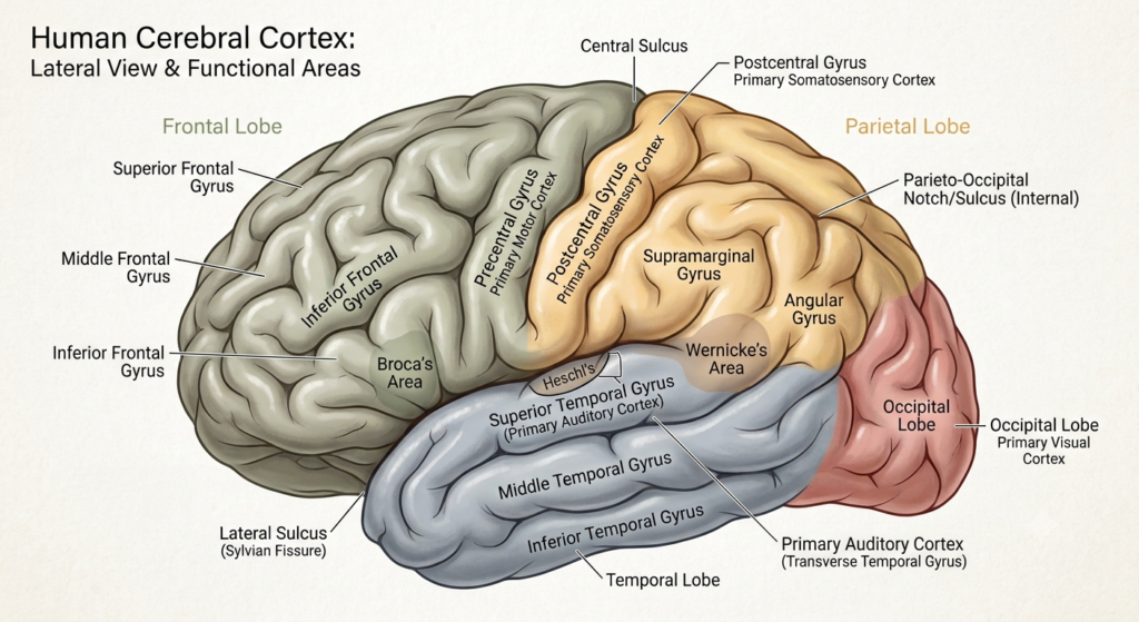
Each hemisphere’s surface is commonly divided into four lobes, and each lobe takes part in several functions rather than just one. The frontal lobe, at the front, contributes to voluntary movement, planning and goal-directed behavior, working memory, attention, holding back inappropriate behavior, decision-making, and speech production. These abilities are spread across several frontal regions rather than run by one isolated executive center.
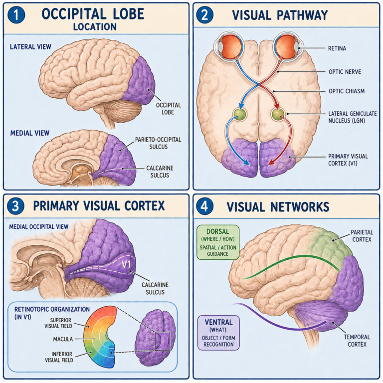
The parietal lobe, behind the frontal lobe, processes touch and other body sensations, awareness of body position, the integration of sensory information, spatial attention, and the sensory guidance of actions. The primary somatosensory cortex sits in its postcentral gyrus. The temporal lobe, below the frontal and parietal lobes, contributes to hearing, language comprehension, recognizing objects and faces, memory, and emotional processing, and memory-related structures including the hippocampal formation lie in its medial part. The occipital lobe, at the back, is mainly associated with vision, and its networks help analyze shape, color, motion, and location.
The insula, or insular cortex, lies hidden deep inside the lateral sulcus, covered by parts of the frontal, parietal, and temporal lobes. It takes part in taste, internal body sensation, pain processing, emotional experience, and autonomic regulation, which is why some classifications call it a fifth lobe. A limbic lobe is also sometimes described on the inner surface, though this is an anatomical grouping rather than a fully independent functional system.
Structures hidden deep inside
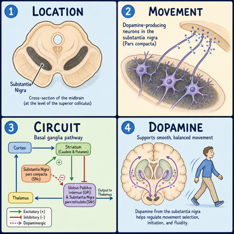
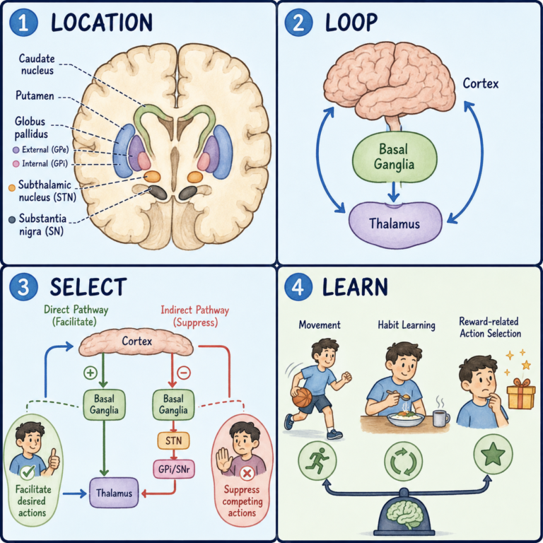
The cerebrum holds more than the cortex and white matter. Deep within each hemisphere are clusters of gray matter. The basal nuclei, usually discussed as part of the basal ganglia, include the caudate nucleus, putamen, and globus pallidus. They take part in circuits for selecting and regulating actions, movement, habit learning, motivation, and reinforcement-based learning. They don’t directly command individual muscles, but influence cortical and brainstem systems through interconnected circuits.
The hippocampal formation, in the medial temporal region, is important for forming and organizing certain kinds of memory and for representing spatial and contextual information. It is not the brain’s only memory structure, since memory depends on distributed networks. The amygdala, in the front of the medial temporal region, helps evaluate biologically and emotionally significant information and learn associations involving threat, reward, and other important events. It is often called the fear center, but that label is too narrow. Each hemisphere also contains a lateral ventricle, a cavity filled with cerebrospinal fluid that connects to the third ventricle through openings called the interventricular foramina.
What the cerebrum does as a whole
No complex ability belongs to one small location. Sensory processing happens in stages: primary sensory areas receive major inputs, and surrounding association areas interpret and combine them. Motor regions of the cortex help plan and produce voluntary actions, sending signals down to the brainstem and spinal cord, while the basal ganglia, cerebellum, thalamus, sensory systems, and spinal circuits provide feedback and modulation.
Language relies on distributed networks, usually with some left-hemisphere dominance, in which frontal, temporal, and parietal regions cooperate during speaking, understanding, reading, and writing. Assigning all speech production to Broca’s area or all comprehension to Wernicke’s area is an oversimplification. For memory, medial temporal structures are especially important for forming certain new memories, while long-term knowledge is stored across widespread cortical networks. Skills, habits, emotional learning, and working memory rely on partly different but interacting systems. Prefrontal and connected networks help people keep goals in mind, compare options, suppress inappropriate responses, solve problems, and adjust to new circumstances. Social behavior draws on frontal, temporal, limbic, sensory, and memory networks together and can’t be pinned to a single personality center.
The cerebrum’s real power lies in this communication. It makes flexible, context-sensitive behavior possible by letting sensation, memory, emotion, and current goals shape action. Even a structurally intact cortical area can work poorly if its white-matter connections or related subcortical circuits are disrupted. And it depends on the rest of the brain: the brainstem supports wakefulness and essential bodily functions, while the cerebellum contributes to coordination and aspects of learning and cognition. The claim that humans use only 10 percent of the cerebrum has no scientific basis. Activity shifts with the task and state, but normal function depends on networks throughout the brain.
When the cerebrum is damaged
The cerebrum can be damaged by stroke, traumatic brain injury, tumors, seizures, infection, inflammation, neurodegenerative disease, or developmental disorders. Possible effects include weakness or impaired movement, loss or alteration of sensation, language problems, loss of part of the visual field, memory difficulty, trouble with attention or planning, changes in motivation or behavior, difficulty recognizing objects, faces, or spatial relationships, and seizures. Symptoms depend on the precise location, extent, and cause of the damage, and on which connecting pathways and wider networks are affected. A symptom can’t always be traced to one isolated spot, so interpreting it requires a neurological assessment and, when appropriate, brain imaging or other tests.
This article is educational background, not a diagnostic guide. Sudden weakness, numbness, vision loss, or trouble speaking needs emergency medical care.
Related: What Is the Cerebral Cortex? Structure and Function · What Is the Brain?
References
- Standring S, Tubbs RS, eds. Gray’s Anatomy: The Anatomical Basis of Clinical Practice. 43rd ed. Elsevier; 2025.
- Kandel ER, Koester JD, Mack SH, Siegelbaum SA, eds. Principles of Neural Science. 6th ed. McGraw Hill; 2021.
- Blumenfeld H. Neuroanatomy Through Clinical Cases. 3rd ed. Oxford University Press; 2021.
- Vanderah TW, Gould DJ. Nolte’s The Human Brain: An Introduction to Its Functional Anatomy. 9th ed. Elsevier; 2025.
- Waxman SG. Clinical Neuroanatomy. 29th ed. McGraw Hill; 2020.
- OpenStax. The Central Nervous System: The Cerebrum. Anatomy and Physiology 2e. 2022.
- Javed K, Reddy V, Lui F. Neuroanatomy, Cerebral Cortex. StatPearls Publishing; updated 2023.
- Bui T, Das JM. Neuroanatomy, Cerebral Hemisphere. StatPearls Publishing; updated 2023.
- National Institute of Neurological Disorders and Stroke. Brain Basics: Know Your Brain. Updated 2025.
Written by: MedMaru Editorial Team
Reviewed for medical accuracy by:
S. Chang, KMD