How Does the Brain Control Human Movement?
How Does the Brain Control Movement?
Introduction
Every voluntary movement—whether reaching for a cup, typing on a keyboard, or taking a single step—depends on the precise coordination of multiple regions of the brain.
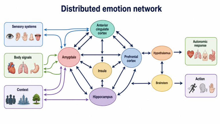
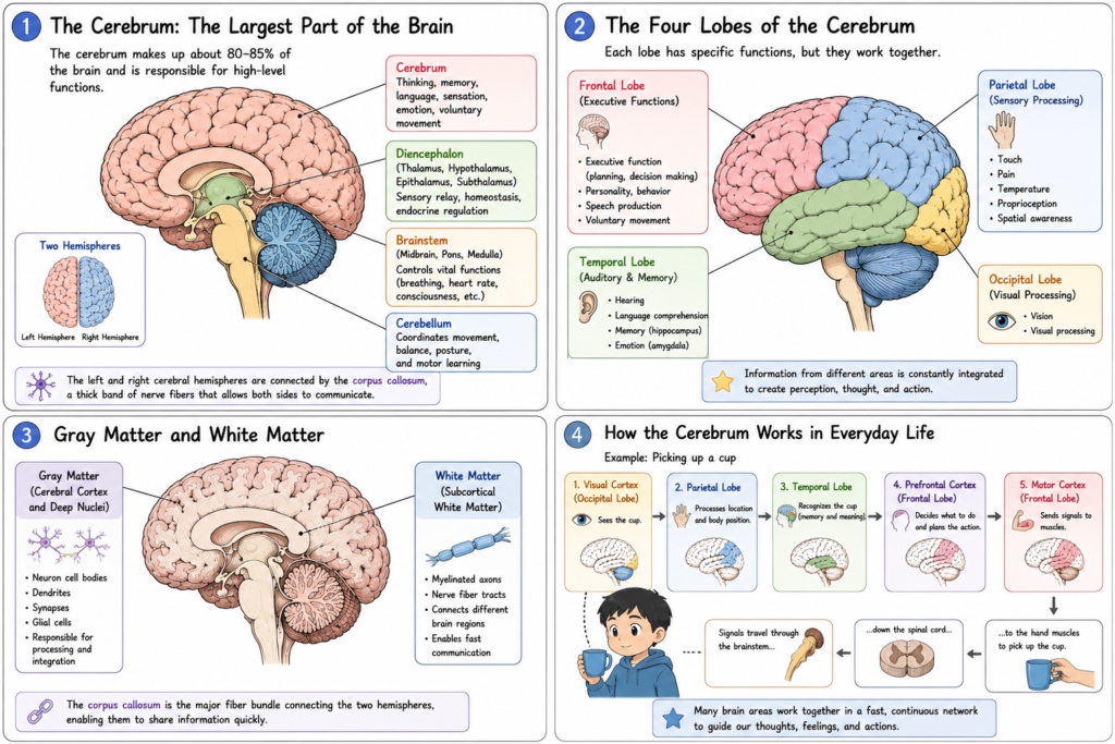
Movement is not controlled by a single “movement center.” Instead, it results from the continuous interaction of the cerebral cortex, basal ganglia, cerebellum, brainstem, spinal cord, and peripheral nerves. Each structure has a specialized role, and together they ensure that movements are accurate, efficient, and adaptable.
Understanding how the brain controls movement provides the foundation for learning neurological disorders such as Parkinson’s disease, Huntington’s disease, cerebellar ataxia, and stroke.
The brain controls movement through a network of interconnected structures. The motor cortex initiates voluntary movement, the basal ganglia help select and regulate movement, the cerebellum coordinates and fine-tunes movement, the brainstem relays and organizes motor signals, and the spinal cord delivers commands to muscles through motor neurons.
How Does the Brain Control Movement?
Voluntary movement follows a highly organized pathway.
A simplified sequence is:
Decision to Move
        ↓
Prefrontal Cortex
        ↓
Premotor Cortex & SMA
        ↓
Primary Motor Cortex
        ↓
Brainstem
        ↓
Spinal Cord
        ↓
Motor Neuron
        ↓
Skeletal Muscle
At every stage, the cerebellum and basal ganglia continuously monitor and adjust the movement.
Step 1. Planning the Movement
Before a movement begins, the brain decides what action should be performed.
The prefrontal cortex is responsible for:
- Goal-directed behavior
- Decision-making
- Motor planning
- Choosing appropriate actions
Once a movement has been selected, the information is transmitted to motor planning areas.
Step 2. Preparing the Movement
Two cortical regions prepare the movement.
Premotor Cortex
The premotor cortex helps prepare movements that depend on external sensory information.
Examples include:
- Catching a ball
- Reaching toward an object
- Responding to visual cues
Supplementary Motor Area (SMA)
The SMA helps organize internally generated movements.
Examples include:
- Walking
- Playing the piano
- Performing a memorized sequence of actions
Together, these regions prepare the motor program before movement begins.
Step 3. Initiating Movement
The primary motor cortex (M1) generates the motor commands.
Neurons in M1 send signals through the corticospinal tract, one of the brain’s major descending motor pathways.
These signals travel toward the spinal cord, where they activate lower motor neurons.
Step 4. Selecting and Regulating Movement
The basal ganglia help determine:
- Which movement should occur
- When movement should begin
- How unwanted movements are suppressed
Rather than generating movement directly, the basal ganglia regulate motor output by communicating with the motor cortex through the thalamus.
Disorders affecting this system include:
- Parkinson disease
- Huntington disease
- Dystonia
Step 5. Coordinating Movement
The cerebellum continuously compares:
- Intended movement
- Actual movement
If differences are detected, the cerebellum rapidly adjusts muscle activity.
This process improves:
- Balance
- Accuracy
- Timing
- Motor learning
Without normal cerebellar function, movements become poorly coordinated and imprecise.
Step 6. Relaying Motor Signals
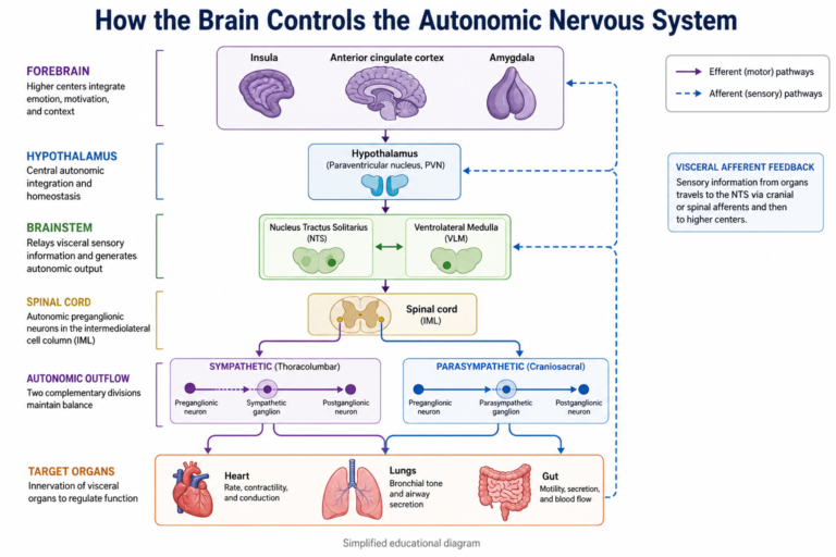
The brainstem serves as the major communication pathway between the brain and spinal cord.
It also contributes to:
- Posture
- Muscle tone
- Eye movements
- Head movements
- Automatic motor control
Several descending motor pathways originate in the brainstem, including the vestibulospinal and reticulospinal tracts.
Step 7. Activating Muscles
Motor commands reach the spinal cord, where upper motor neurons synapse with lower motor neurons.
Lower motor neurons send electrical signals through peripheral nerves to skeletal muscles.
At the neuromuscular junction, acetylcholine is released, causing muscle fibers to contract.
This is the final step that produces visible movement.
Sensory Feedback
Movement does not end once muscles contract.
Sensory receptors continuously provide feedback, including:
- Proprioceptors
- Muscle spindles
- Golgi tendon organs
- Joint receptors
- Skin mechanoreceptors
This information travels back to the spinal cord, cerebellum, and cerebral cortex, allowing movements to be corrected in real time.
Without sensory feedback, movements become inaccurate and unstable.
Why Brain Control of Movement Is Important
Normal movement depends on the cooperation of multiple brain regions.
Each structure contributes a unique role:
- Prefrontal cortex → Decides what to do.
- Premotor cortex & SMA → Prepare the movement.
- Primary motor cortex → Sends motor commands.
- Basal ganglia → Select and regulate movement.
- Cerebellum → Coordinates movement.
- Brainstem → Relays and organizes motor pathways.
- Spinal cord → Activates muscles.
Damage to any one of these regions produces different patterns of movement disorders.
Clinical Relevance
Neurological diseases affecting motor control include:
- Parkinson disease
- Huntington disease
- Stroke
- Amyotrophic lateral sclerosis (ALS)
- Multiple sclerosis
- Cerebellar ataxia
- Spinal cord injury
Each disorder affects different parts of the motor system and therefore produces distinct clinical symptoms.
Common Misunderstandings
“The motor cortex alone controls movement.”
Incorrect.
Movement depends on continuous interaction among the motor cortex, basal ganglia, cerebellum, brainstem, spinal cord, and sensory feedback systems.
“The cerebellum starts movement.”
Incorrect.
The cerebellum does not initiate voluntary movement. Instead, it coordinates and fine-tunes movements that have already been planned.
“The basal ganglia send signals directly to muscles.”
Incorrect.
The basal ganglia influence movement indirectly through circuits involving the thalamus and cerebral cortex.
- Movement is controlled by multiple interconnected brain regions.
- The motor cortex initiates voluntary movement.
- The basal ganglia regulate movement selection.
- The cerebellum coordinates and refines movement.
- The brainstem and spinal cord transmit motor commands to muscles.
- Sensory feedback continuously adjusts movement for accuracy.
Related Articles
References
- Neuroanatomy, Motor Neuron. StatPearls. NCBI Bookshelf.
- Jimsheleishvili S, Dididze M. Neuroanatomy, Cerebellum. StatPearls. NCBI Bookshelf.
- Manto M, et al. Consensus paper: roles of the cerebellum in motor control. Cerebellum. 2012;11(2):457-487. doi:10.1007/s12311-011-0331-9.
Written by: MedMaru Editorial Team
Reviewed for medical accuracy by:
S. Chang, KMD