Brain Ventricles: Location, Function, and Anatomy
The brain ventricles are four connected, fluid-filled chambers inside the brain where cerebrospinal fluid is produced and through which it circulates. Cerebrospinal fluid, or CSF, is the clear liquid that fills the ventricles and surrounds the brain and spinal cord. The ventricles work like the brain’s plumbing. Fluid is made inside the chambers, flows through connecting passages in one general direction, and eventually drains out to bathe the whole brain and spinal cord in a protective cushion.
Four chambers, one system
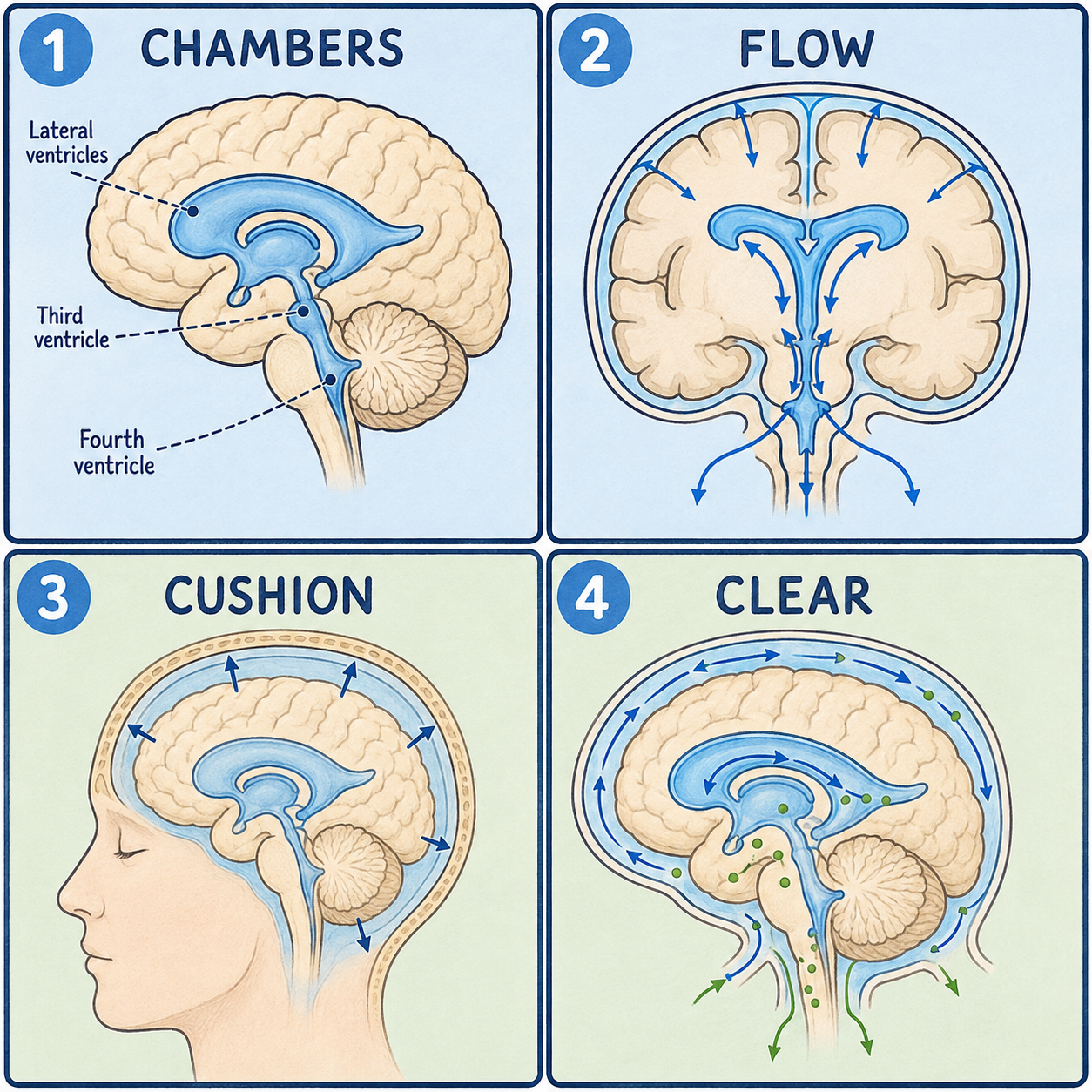
There are four ventricles, and they form one continuous system rather than four separate pools. The two lateral ventricles are the largest, one inside each cerebral hemisphere. The third ventricle is a narrow, slit-like chamber in the midline, between the two halves of the thalamus. The fourth ventricle is a diamond-shaped chamber between the brainstem and the cerebellum, and it is the lowest of the four.
Most CSF is produced by the choroid plexus, a specialized tissue rich in blood vessels that lines parts of the ventricles. The ependymal cells lining the ventricle walls make a smaller contribution.
How the fluid moves
The chambers connect in sequence. Each lateral ventricle opens into the third ventricle through the interventricular foramen, also called the foramen of Monro. The third ventricle connects to the fourth through the cerebral aqueduct, a narrow canal running through the midbrain. The fourth ventricle sits with the pons and medulla oblongata in front of it and the cerebellum behind.
From the fourth ventricle, CSF leaves the ventricular system through three openings: two lateral apertures, the foramina of Luschka, and one median aperture, the foramen of Magendie. They lead into the subarachnoid space, the fluid-filled layer surrounding the entire brain and spinal cord beneath the meninges. A small amount also continues down the central canal, a thin channel inside the spinal cord.
Picture the system as a small branching river: two headwater streams, the lateral ventricles, merge into one channel that empties out at the bottom. Unlike a real river, though, CSF circulation is continuously replenished. Fluid is constantly being made and constantly being reabsorbed, so the river never runs dry. That also corrects a common assumption that CSF simply sits still around the brain. It is always on the move.
As it circulates, CSF cushions the brain against shocks, helps keep the chemical environment around neurons stable, supplies nutrients, and carries some immune cells, before eventually being reabsorbed into the bloodstream. How that reabsorption happens is described in more detail in the meninges article.
When the flow gets blocked
Because the ventricles form a single connected pathway, a blockage anywhere along it can back up CSF like a clog in a pipe. The resulting buildup is called hydrocephalus, literally “water on the brain,” and it raises pressure inside the skull.
There are two broad ways this happens. In obstructive hydrocephalus, a tumor or cyst blocks one of the narrow connecting passages. The chambers upstream of the blockage enlarge because fluid keeps being produced with nowhere to go. The cerebral aqueduct, being so narrow, is one vulnerable point. A blockage right at the outlets of the fourth ventricle, the most downstream chamber, can cause the entire ventricular system above it to enlarge. In communicating hydrocephalus, the passages are open, but CSF is not reabsorbed properly further downstream.
So hydrocephalus does not usually mean the brain is making too much fluid. Many cases result from a blocked pathway or a problem with reabsorption rather than overproduction.
This article is educational background, not a diagnostic guide. A severe headache with vomiting, worsening drowsiness, changes in vision or walking, or, in infants, a rapidly enlarging head or bulging soft spot needs prompt medical evaluation.
Related: What Is the Cerebral Cortex? · What Is the Brain? · What Is the Cerebrum?
References
- StatPearls. “Neuroanatomy, Fourth Ventricle.” NCBI Bookshelf, NBK546577 — https://www.ncbi.nlm.nih.gov/books/NBK546577/
Written by: MedMaru Editorial Team
Reviewed for medical accuracy by:
S. Chang, KMD