Cerebrum: Anatomy, Lobes, and Function
What Is the Cerebrum? Structure and Function
Introduction
The cerebrum is the largest part of the human brain. It forms the prominent, folded structure visible when the brain is viewed from above or from the side.
The cerebrum allows us to perceive the world, produce voluntary movements, use language, form memories, regulate behavior, and perform complex mental tasks. However, it does not perform these functions alone. It works continuously with the thalamus, brainstem, cerebellum, spinal cord, and other parts of the nervous system.
Understanding the cerebrum provides a foundation for learning about the cerebral cortex, the four cerebral lobes, the basal ganglia, and many other brain structures.
The cerebrum is the largest part of the brain and consists mainly of the right and left cerebral hemispheres. Each hemisphere contains an outer cerebral cortex, underlying white matter, and deep collections of gray matter.
The cerebrum contributes to:
- Conscious sensation
- Voluntary movement
- Language
- Learning and memory
- Attention
- Planning and decision-making
- Emotion and motivation
- Recognition of objects, faces, and spatial relationships
These functions arise from networks distributed across the cerebrum and connected with other parts of the nervous system.
What Is the Cerebrum?
The cerebrum is the large upper portion of the brain formed by the two cerebral hemispheres. During embryonic development, the cerebral hemispheres arise from the telencephalon, a division of the developing forebrain.
The term cerebrum should not be used as a synonym for the entire brain. The brain also includes other major structures, such as the:
- Diencephalon, including the thalamus and hypothalamus
- Brainstem
- Cerebellum
The boundaries and terminology used for deep forebrain structures can vary among anatomy texts. In general, however, each cerebral hemisphere includes the cerebral cortex, underlying white matter, and deep gray-matter structures such as the basal nuclei.
What Are the Two Cerebral Hemispheres?
The cerebrum is divided into a left hemisphere and a right hemisphere. A deep groove called the longitudinal fissure separates the hemispheres along most of their upper surface.
The hemispheres are not completely isolated. They communicate through bundles of nerve fibers known as commissural fibers. The largest of these connections is the corpus callosum.
Each hemisphere receives and processes information from both sides of the body, although many important sensory and motor pathways cross from one side to the other. As a result, the left cerebral hemisphere has a strong influence on movement and sensation on the right side of the body, and the right hemisphere has a strong influence on the left side.
Some functions show partial hemispheric specialization. For example, language is predominantly supported by the left hemisphere in most people, while some aspects of spatial attention are more strongly associated with right-hemisphere networks. This division is not absolute. Both hemispheres participate in most complex behaviors and communicate extensively.
What Is the Cerebral Cortex?
The cerebral cortex is the outer layer of neural tissue covering the cerebral hemispheres. It is composed of gray matter containing neuronal cell bodies, dendrites, synapses, supporting glial cells, and blood vessels.
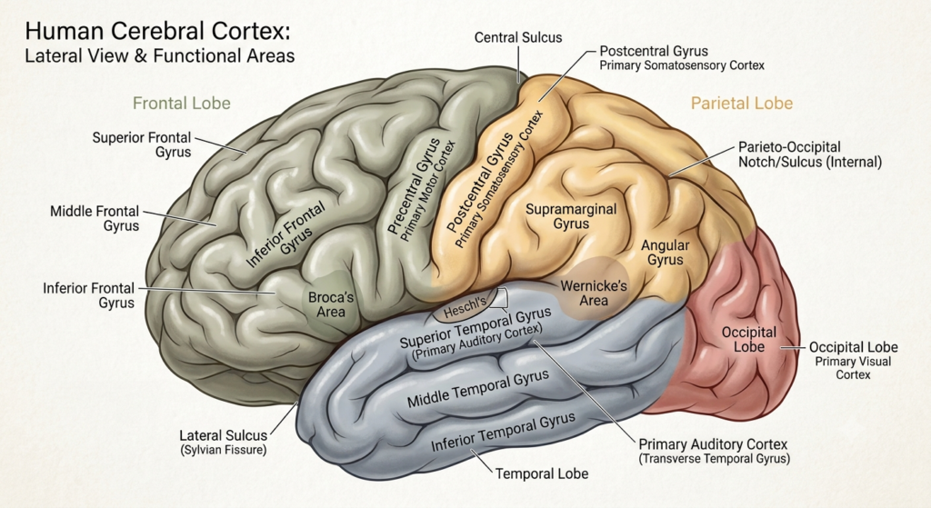
The cortex is highly folded. Its raised ridges are called gyri, and its grooves are called sulci. Particularly deep grooves may be called fissures.
Folding greatly increases the cortical surface area that can fit within the skull. These folds also create recognizable anatomical landmarks that help identify the different lobes and functional regions.
The cerebral cortex is involved in conscious perception, voluntary movement, language, memory, attention, and many other complex functions. It should not, however, be viewed as a single control layer acting independently. Cortical functions depend on constant communication with deeper cerebral structures, the thalamus, basal ganglia, cerebellum, and brainstem.
What Is Cerebral White Matter?
Beneath the cerebral cortex lies cerebral white matter. Its lighter appearance results mainly from the myelin surrounding many nerve fibers.
These fibers carry signals between different parts of the nervous system. They can be broadly grouped into three types:
- Association fibers connect cortical regions within the same hemisphere.
- Commissural fibers connect the two hemispheres.
- Projection fibers connect the cerebral cortex with deeper structures, the brainstem, and spinal cord.
The corpus callosum is the largest commissural pathway. The internal capsule is an important concentration of projection fibers carrying sensory and motor signals.
White matter is therefore not simply passive tissue beneath the cortex. It forms the communication system that allows distributed brain regions to operate as coordinated networks.
What Are the Lobes of the Cerebrum?
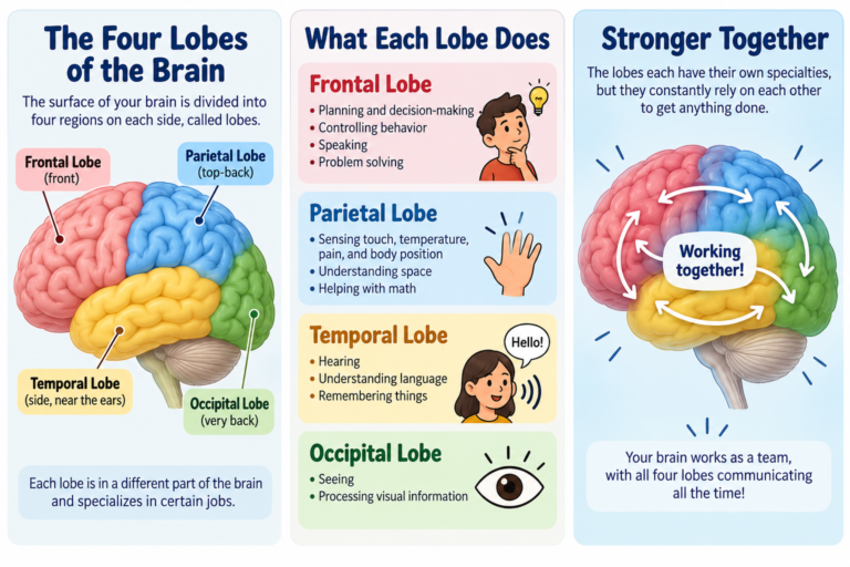
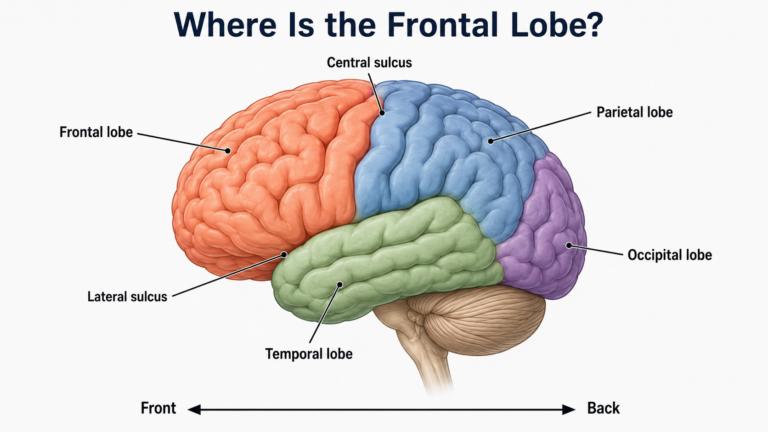
The surface of each cerebral hemisphere is commonly divided into four major lobes.
Frontal Lobe
The frontal lobe is located toward the front of the cerebrum. Its networks contribute to:
- Voluntary movement
- Planning and goal-directed behavior
- Working memory
- Attention
- Behavioral inhibition
- Decision-making
- Speech production
These abilities are distributed across several frontal regions rather than controlled by one isolated “executive center.”
Parietal Lobe
The parietal lobe is located behind the frontal lobe. It contributes to:
- Processing touch and other bodily sensations
- Awareness of body position
- Integration of sensory information
- Spatial attention
- Guidance of actions using sensory information
The primary somatosensory cortex is located in the postcentral gyrus of the parietal lobe.
Temporal Lobe
The temporal lobe lies below the frontal and parietal lobes. It participates in:
- Hearing
- Language comprehension
- Recognition of objects and faces
- Memory
- Emotional processing
Important memory-related structures, including the hippocampal formation, are located in the medial temporal region.
Occipital Lobe
The occipital lobe is located at the back of the cerebrum and is primarily associated with vision.
Visual signals reaching the cortex are processed through multiple occipital and connected cortical regions. These networks help analyze features such as shape, color, motion, and spatial location.
What About the Insula?
The insula, or insular cortex, lies deep within the lateral sulcus and is covered by portions of the frontal, parietal, and temporal lobes.
It participates in processes including:
- Taste
- Internal bodily sensation
- Pain processing
- Emotional experience
- Autonomic regulation
Some anatomical classifications therefore describe the insula as a fifth cerebral lobe. A “limbic lobe” may also be described on the medial surface, although this is an anatomical grouping rather than a completely independent functional system.
What Structures Lie Deep Within the Cerebrum?
The cerebrum contains more than the visible cortex and underlying white matter. Collections of gray matter are also located deep within the hemispheres.
Basal Nuclei
The basal nuclei, commonly discussed as part of the basal ganglia, include the caudate nucleus, putamen, and globus pallidus. They participate in circuits involved in:
- Selecting and regulating actions
- Movement
- Habit learning
- Motivation
- Reinforcement-based learning
They do not directly command individual muscles. Instead, they influence cortical and brainstem systems through interconnected neural circuits.
Hippocampal Formation
The hippocampal formation lies in the medial temporal region. It is important for forming and organizing certain types of memory and for representing spatial and contextual information.
It is not the brain’s only memory structure. Memory depends on distributed networks involving numerous cortical and subcortical regions.
Amygdala
The amygdala is located in the anterior medial temporal region. It contributes to evaluating biologically and emotionally significant information and to learning associations involving threat, reward, and other important events.
It is often called the brain’s “fear center,” but this description is too narrow. Its functions extend beyond fear and depend on connections with many other regions.
Lateral Ventricles
Each cerebral hemisphere contains a lateral ventricle, a cavity filled with cerebrospinal fluid. The lateral ventricles are part of the ventricular system and communicate with the third ventricle through the interventricular foramina.
What Does the Cerebrum Do?
The cerebrum contributes to an exceptionally wide range of functions, but no complex ability belongs to only one small location.
Sensory Processing
Cerebral networks help transform signals from the eyes, ears, skin, muscles, joints, and other sensory systems into meaningful perceptions.
Sensory processing occurs in stages. Primary sensory areas receive major inputs, while surrounding and interconnected association areas help interpret and combine information.
Voluntary Movement
Motor regions of the cerebral cortex contribute to planning and producing voluntary actions. Their signals reach the brainstem and spinal cord through descending pathways.
Movement also depends on feedback and modulation from the basal ganglia, cerebellum, thalamus, sensory systems, and spinal circuits.
Language
Language relies on distributed networks, usually with a degree of left-hemisphere dominance. Frontal, temporal, and parietal regions work together during speech production, comprehension, reading, and writing.
Descriptions that assign all language production to Broca’s area or all comprehension to Wernicke’s area are overly simplified.
Memory and Learning
The cerebrum supports several kinds of learning and memory. Medial temporal structures are particularly important for forming certain new memories, while long-term knowledge is represented across widespread cortical networks.
Skills, habits, emotional learning, and working memory depend on partly different but interacting systems.
Executive and Social Functions
Prefrontal and connected networks help people maintain goals, compare options, suppress inappropriate responses, solve problems, and adjust behavior according to changing circumstances.
Social behavior also requires interaction among frontal, temporal, limbic, sensory, and memory networks. It cannot be assigned to a single “personality center.”
Why Is the Cerebrum Important?
The cerebrum makes flexible, context-sensitive behavior possible. It allows sensory information, memory, emotion, and current goals to influence action.
Its importance comes not only from the functions of individual regions but also from communication among them. Even a structurally intact cortical area may function poorly if its white-matter connections or related subcortical circuits are disrupted.
The cerebrum also depends on other brain divisions. For example, the brainstem supports wakefulness and essential physiological functions, while the cerebellum contributes to movement coordination and aspects of learning and cognition.
Clinical Relevance
Damage to the cerebrum can result from stroke, traumatic brain injury, tumors, seizures, infection, inflammation, neurodegenerative disease, or developmental disorders.
Possible effects include:
- Weakness or impaired voluntary movement
- Loss or alteration of sensation
- Language impairment
- Visual field loss
- Memory difficulty
- Problems with attention or planning
- Changes in motivation or behavior
- Difficulty recognizing objects, faces, or spatial relationships
- Seizures
Symptoms depend on the precise location, extent, and cause of the damage. They also depend on which connecting pathways and broader networks are affected.
A symptom cannot always be traced to one isolated cerebral location. Clinical interpretation requires neurological assessment and, when appropriate, brain imaging or other tests.
Common Misunderstandings About the Cerebrum
“The Cerebrum and the Brain Are the Same Thing”
The cerebrum is the largest part of the brain, but the brain also includes the diencephalon, brainstem, and cerebellum.
“The Left Brain Is Logical and the Right Brain Is Creative”
Some functions show hemispheric specialization, but complex thinking and creativity recruit networks in both hemispheres. People cannot be scientifically classified as strictly “left-brained” or “right-brained.”
“Each Lobe Has Only One Function”
Every cerebral lobe participates in multiple functions. Most meaningful behaviors depend on networks extending across several lobes and deeper structures.
“Gray Matter Does the Thinking, While White Matter Is Just Wiring”
Gray matter contains many sites of neural processing, while white matter enables communication among them. Both are essential to cerebral function.
“Humans Use Only 10 Percent of the Cerebrum”
There is no scientific basis for the claim that most of the cerebrum remains unused. Different regions vary in activity according to the task and state, but normal brain function depends on networks distributed throughout the brain.
Related Articles
- What Is the Cerebral Cortex? Structure and Function
- What Are the Major Parts of the Brain? A Complete Guide to Brain Anatomy
- What Is the Brain? Definition, Structure, and Function
References
- Standring S, Tubbs RS, eds. Gray’s Anatomy: The Anatomical Basis of Clinical Practice. 43rd ed. Elsevier; 2025.
- Kandel ER, Koester JD, Mack SH, Siegelbaum SA, eds. Principles of Neural Science. 6th ed. McGraw Hill; 2021.
- Blumenfeld H. Neuroanatomy Through Clinical Cases. 3rd ed. Oxford University Press; 2021.
- Vanderah TW, Gould DJ. Nolte’s The Human Brain: An Introduction to Its Functional Anatomy. 9th ed. Elsevier; 2025.
- Waxman SG. Clinical Neuroanatomy. 29th ed. McGraw Hill; 2020.
- OpenStax. The Central Nervous System: The Cerebrum. Anatomy and Physiology 2e. 2022.
- Javed K, Reddy V, Lui F. Neuroanatomy, Cerebral Cortex. StatPearls Publishing; updated 2023.
- Bui T, Das JM. Neuroanatomy, Cerebral Hemisphere. StatPearls Publishing; updated 2023.
- National Institute of Neurological Disorders and Stroke. Brain Basics: Know Your Brain. Updated 2025.
Written by: MedMaru Editorial Team
Reviewed for medical accuracy by:
S. Chang, KMD