What Is Excitation–Inhibition Balance in the Brain?
“Balance” Doesn’t Mean Equal Amounts
It’s tempting to picture excitation-inhibition balance as a scale with equal weight on both sides — half the signals pushing neurons to fire, half holding them back. That picture is wrong, and it matters that it’s wrong: real circuits don’t aim for a fixed 50/50 ratio anywhere in the brain. What they maintain is a constantly shifting coordination between signals that promote activity and signals that regulate it, adjusted in timing and strength as demands change moment to moment. A circuit counts as functionally balanced when it can respond strongly to something meaningful without spiraling into runaway activity — not when its excitatory and inhibitory inputs happen to match numerically.
The same coordination, at every scale
This balancing act isn’t a single brain-wide property — it’s replicated at several nested levels. At the synapse, a single neuron can receive thousands of excitatory and inhibitory inputs simultaneously, and their location, strength, and timing all shape whether that neuron ultimately fires. Zoom out one level, and the same logic plays out inside local circuits: in the cerebral cortex, excitatory pyramidal neurons communicate constantly with inhibitory interneurons, which regulate their neighbors through both feedback and feedforward inhibitory loops. Parvalbumin-expressing interneurons, in particular, are central to fast inhibition and to keeping cortical activity properly timed and synchronized. Zoom out further still, and excitation and inhibition shape how entire brain regions coordinate with each other — sensory processing, movement, attention, memory, and the rhythmic oscillations that show up in EEG recordings all depend on this same interplay operating at network scale.
Glutamate and GABA aren’t the whole story
Glutamate is the principal excitatory neurotransmitter in the central nervous system, acting through AMPA, NMDA, and metabotropic receptors. GABA is the principal inhibitory neurotransmitter in the mature brain, acting mainly through GABA-A and GABA-B receptors. That’s a useful shorthand, and also an incomplete one: a neurotransmitter’s effect depends on which receptor it binds and the ion gradients across that particular neuron’s membrane, not on the molecule alone. GABA is a clear illustration — during early development, it can actually be depolarizing rather than inhibitory, even while still shaping circuit activity in complex ways. It also follows that measuring total glutamate and GABA in a region doesn’t tell you the functional E-I balance of the circuits there; concentration, receptor activity, synaptic strength, neuronal excitability, and connectivity are all separate dimensions of the same system, and they don’t move in lockstep.
How circuits keep themselves from running away
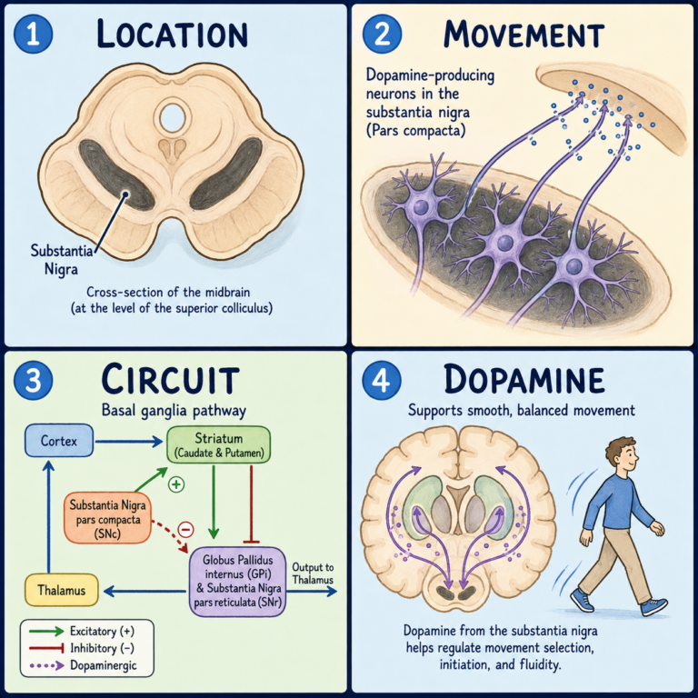
Several distinct mechanisms cooperate to hold this system within a workable range. Feedback inhibition kicks in when excitatory neurons activate the inhibitory interneurons that then suppress further firing in that same circuit. Feedforward inhibition works slightly differently — an incoming signal activates both a principal neuron and an inhibitory neuron at once, with the inhibitory neuron constraining the timing or duration of the principal neuron’s response. Longer-term, synaptic plasticity lets both excitatory and inhibitory connections change strength as circuits adapt to learning and experience, while homeostatic plasticity acts as a stabilizer underneath that: if a neuron or circuit drifts too active or too quiet, compensatory changes in synaptic strength, receptor expression, or intrinsic excitability pull it back, without erasing the learning-related changes that got it there. Neuromodulators — dopamine, acetylcholine, serotonin, norepinephrine — add another layer on top, shifting neuronal excitability and synaptic effectiveness according to attention, arousal, sleep, and behavioral state. E-I balance, in other words, isn’t a fixed setting; it moves with what the brain is doing.
Inhibition isn’t just an “off” switch
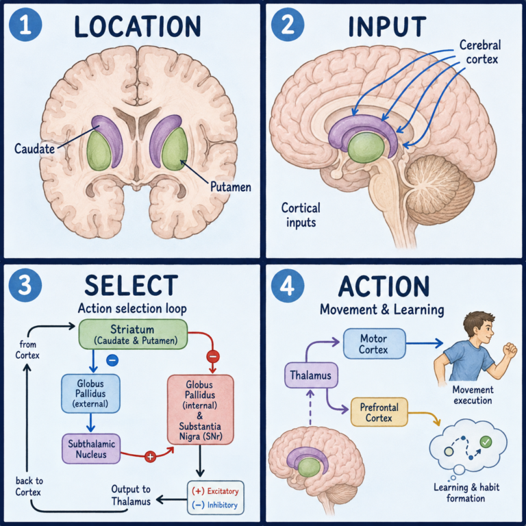
It’s worth being explicit about what inhibition actually accomplishes, because “restraining activity” undersells it. Inhibition sharpens responses, controls the timing of firing, helps select among competing signals, and synchronizes groups of neurons — it’s doing active computational work, not just damping things down. And more inhibition isn’t automatically better: circuits still need enough excitation to transmit information and to change in response to experience. The system needs both, coordinated correctly, not one dialed up and the other dialed down.
When the coordination breaks down
A substantial loss of inhibitory restraint, or excessive recurrent excitation, can push circuits into hypersynchronous, uncontrolled activity — epilepsy is the clearest clinical example of this kind of breakdown. E-I-related changes have also been studied in autism, schizophrenia, and Alzheimer’s disease, among other conditions, but it’s worth being precise about what that means: these conditions are biologically diverse, their symptoms aren’t reducible to a single brain-wide E-I ratio, and in most cases it isn’t clear whether an observed E-I change is a primary cause, a downstream consequence of something else, a compensatory response, or a feature specific to particular cells, circuits, or developmental windows. “E-I imbalance” functions as a useful circuit-level framework for asking questions — it isn’t a diagnosis, and no currently established clinical test can diagnose a complex neurodevelopmental or psychiatric condition by measuring a single E-I ratio.
Measuring something you can’t observe directly
There’s no single test that captures the brain’s overall E-I balance, because there isn’t one biological quantity to capture. Researchers reach for different tools depending on what they’re after: intracellular and patch-clamp recordings for excitatory and inhibitory currents directly in tissue, EEG or MEG for population-level electrical activity, transcranial magnetic stimulation, magnetic resonance spectroscopy for regional glutamate and GABA concentrations, molecular assays of receptors and neurotransmitters, and computational models that simulate circuit behavior. Each of these captures a different feature or indirect marker of the system — magnetic resonance spectroscopy, for instance, estimates neurotransmitter concentration but says nothing directly about moment-to-moment synaptic excitation and inhibition. Results from different methods shouldn’t be treated as measuring the same underlying thing, because in a meaningful sense, they aren’t.
Related: What Are Excitatory and Inhibitory Signals? · How Do Neurons Combine Excitatory and Inhibitory Signals? · What Are Neurotransmitters?
References
- Tatti R, Haley MS, Swanson OK, Tselha T, Maffei A. Neurophysiology and Regulation of the Balance Between Excitation and Inhibition in Neocortical Circuits. Biological Psychiatry. 2017;81(10):821-831.
- He HY, Cline HT. What Is Excitation/Inhibition and How Is It Regulated? A Case of the Elephant and the Wisemen. Journal of Experimental Neuroscience. 2019;13:1179069519859371.
- Chen L, Li X, Tjia M, Thapliyal S. Homeostatic Plasticity and Excitation-Inhibition Balance: The Good, the Bad, and the Ugly. Current Opinion in Neurobiology. 2022;75:102553.
- Turrigiano G. Homeostatic Synaptic Plasticity: Local and Global Mechanisms for Stabilizing Neuronal Function. Cold Spring Harbor Perspectives in Biology. 2012;4(1):a005736.
- Okur Z, Schlauri N, Bitsikas V, et al. Control of Neuronal Excitation-Inhibition Balance by BMP-SMAD1 Signalling. Nature. 2024;629(8011):402-409.
Written by: MedMaru Editorial Team
Reviewed for medical accuracy by:
S. Chang, KMD