Occipital Lobe: Location, Function, and Anatomy
The occipital lobe forms the back part of each cerebral hemisphere, and it contains the brain’s primary visual cortex. It receives highly organized visual input, then works with temporal, parietal, frontal, and deeper subcortical networks to turn that input into useful perception and visually guided action. It is the first stop for vision in the cortex, but not the place where seeing is finished.
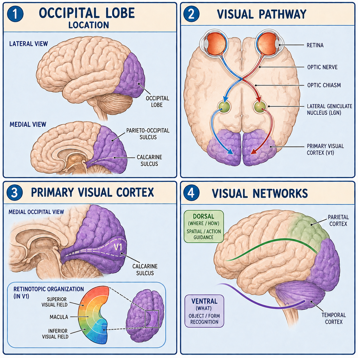
Finding the back of the brain
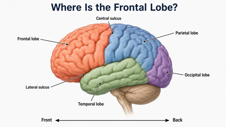
The occipital lobe lies behind the parietal and temporal lobes and above the cerebellum. On the inner, or medial, surface of the brain its front edge is easy to find, because a groove called the parieto-occipital sulcus marks it clearly. On the outer surface the boundary is less obvious. It is usually approximated by drawing a line from the parieto-occipital sulcus down to a small dent called the preoccipital notch. The grooves and ridges on the outer occipital surface vary considerably from person to person, so a fixed three-ridge pattern sometimes shown in diagrams should not be treated as universal.
The most important landmark is on the medial surface. The calcarine sulcus is a groove whose banks hold the primary visual cortex, known as V1 or Brodmann area 17. Above the sulcus lies the cuneus, and below it lies the lingual gyrus. V1 is retinotopically organized, which means it holds an orderly map of what the eyes see. Neighboring points in the visual field map to neighboring patches of cortex, much like neighboring streets sit side by side on a city map. Unlike a fair map, though, this one is distorted: the central part of vision gets a disproportionately large share of cortex near the occipital pole, the very back tip of the lobe.
How signals from the eyes get here
A common assumption is that the occipital lobe is the first part of the brain to receive visual information. It isn’t. The signal takes a journey first. Axons from retinal ganglion cells, the output cells of the retina, bundle together to form the optic nerves. At the optic chiasm, fibers from the inner, nasal half of each retina cross to the other side, while fibers from the outer, temporal half stay on the same side.
The result of that partial crossing is elegant. Each optic tract behind the chiasm carries information about the opposite half of the visual field, from both eyes. Most of these fibers then connect in the lateral geniculate nucleus of the thalamus. From there, white-matter fibers called the optic radiations fan back to V1 around the calcarine sulcus. So one occipital lobe does not handle one eye. Each one handles the opposite half of the world as seen by both eyes. This is also why injury behind the optic chiasm tends to affect the same side of the visual field in both eyes rather than one eye alone.
Seeing takes a network, not a single ladder
V1 performs early analysis of features such as contrast, orientation, and position in space. From there, visual information engages many interconnected areas beyond V1, called extrastriate areas. It does not climb a simple ladder from one area to the next.
Two broad networks are often described. The dorsal network extends toward the parietal cortex and contributes to spatial relationships, motion, attention, and visually guided action. It is often summarized as the where or how pathway. The ventral network extends toward the temporal cortex and contributes to recognizing objects, shapes, colors, words, and faces. It is often summarized as the what pathway. These labels are useful summaries, but they are not the whole story. Both streams contain several interacting pathways, they support overlapping computations, and they exchange information with each other and with frontal and subcortical systems. So the occipital lobe does not create a complete picture by itself. V1 is essential for early cortical vision, yet conscious visual perception depends on networks reaching well beyond the occipital lobe.
What occipital injury can look like
Because the visual field is mapped so precisely, damage here often produces predictable patterns. Injury to the primary visual cortex on one side causes contralateral homonymous hemianopia, which is loss of the same half of the visual field, on the opposite side, in both eyes. Damage limited to the upper or lower bank of the calcarine sulcus causes a quarter-field loss, called quadrantanopia, depending on which part of the map is involved. When visual cortex or the pathways behind the lateral geniculate nucleus are damaged on both sides, a person can lose vision even though the eyes themselves are structurally intact.
Other patterns are less about blindness and more about recognition. Damage to the occipitotemporal visual network can make it hard to recognize objects, faces, colors, or written words, depending on where the lesion is. Seizure activity in an occipital network can cause brief, simple visual experiences such as flashes, colored shapes, or symptoms in part of the visual field.
One nuance deserves attention. Some occipital lesions cause homonymous hemianopia but leave central vision intact, a pattern called macular sparing. It happens when the cortex at the occipital pole that represents the macula, the center of the retina, keeps working. Variable collateral blood supply can protect that tissue after a posterior cerebral artery is blocked, and in some people this includes contributions from branches of the middle cerebral artery. Incomplete lesions and other anatomical variations can matter too. Macular sparing should therefore not be explained as an invariable dual-blood-supply rule.
This article is educational background, not a diagnostic guide. Sudden loss of vision or of part of the visual field needs urgent medical evaluation.
Related: What Is the Cerebral Cortex? · What Is the Brain? · What Is the Cerebrum?
References
- NCBI Bookshelf: Neuroanatomy, Occipital Lobe
- NCBI Bookshelf: The Primary Visual Cortex
- The Mechanism of Macular Sparing
Medically reviewed by S. Chang, KMD.
Written by: MedMaru Editorial Team
Reviewed for medical accuracy by:
S. Chang, KMD