How the Corpus Callosum Connects the Brain’s Two Hemispheres
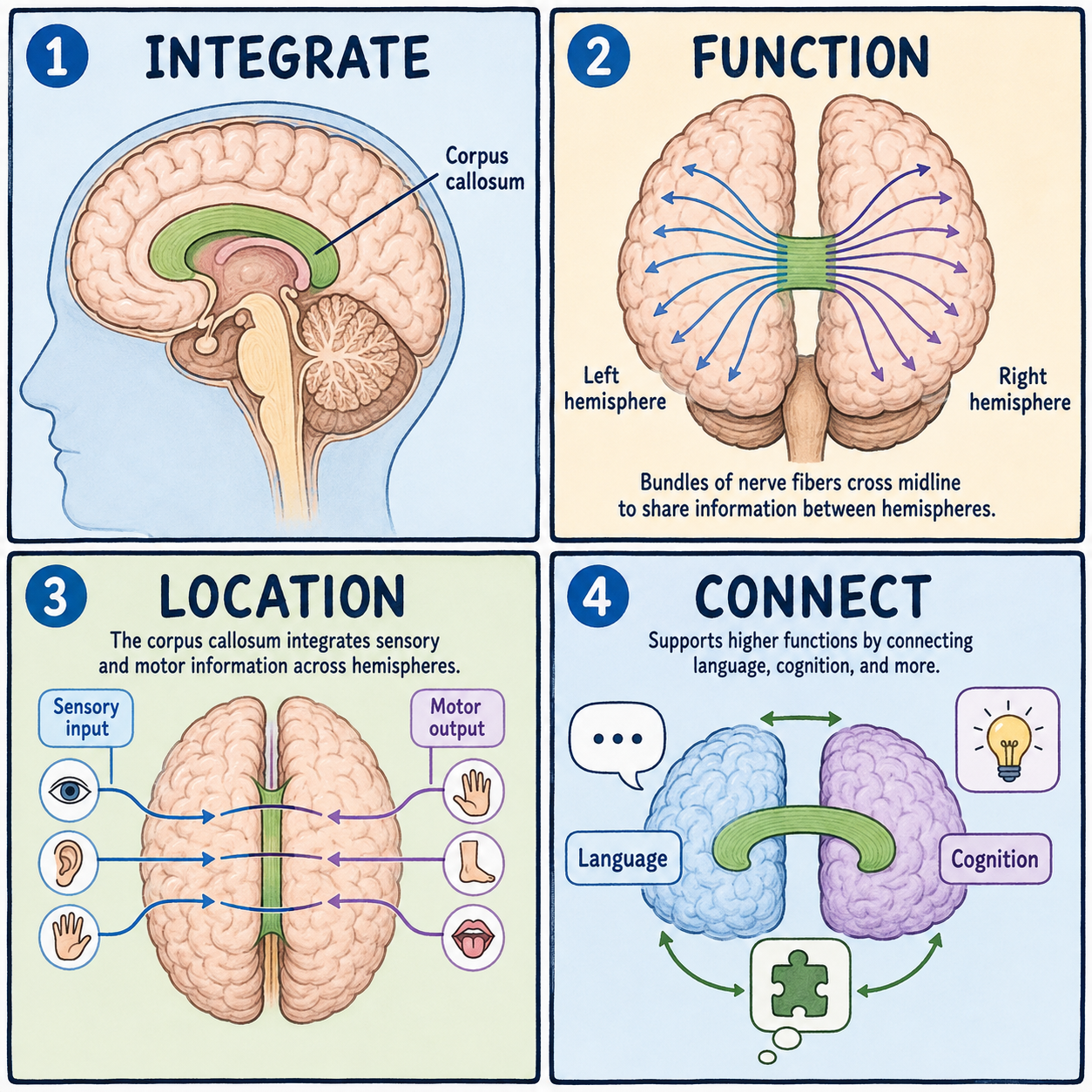
The brain is really two halves working as one team, and the corpus callosum is what lets them talk to each other. It is a thick cable of roughly 200 million nerve fibers connecting the left and right sides. Signals cross it in a fraction of a second, so each side can share what it sees, feels, and plans almost as if the brain were a single unit. When this cable is thin, damaged, or missing, the two hemispheres can’t compare notes properly. That tends to show up as specific gaps in coordination rather than a general loss of thinking ability.
The brain’s largest white-matter bridge
The corpus callosum is the largest white-matter structure in the human brain. White matter is tissue made of long nerve fibers wrapped in myelin, a fatty insulating sheath that makes it paler than the gray matter, where nerve cell bodies are concentrated. The corpus callosum sits deep in the middle of the brain. It arches like a curved bridge above the lateral ventricles and below the longitudinal fissure, the deep groove that separates the left and right cerebral hemispheres.
Picture the two hemispheres as two people at neighboring desks who share one thick phone line. Each can think and act somewhat independently, but the line lets them constantly check in, compare what they are looking at, and coordinate a joint decision. The picture is imperfect, because the hemispheres are not two separate minds, but it captures why the connection matters.
The corpus callosum is not the only bridge across the midline. The anterior commissure is a much smaller fiber bundle that also crosses over. Its main job, though, is connecting smaller, more specific regions such as parts of the temporal lobes, not the broad cortex-to-cortex traffic the corpus callosum carries.
Organized from front to back
From front to back, the corpus callosum is divided into named sections: the rostrum, the genu, which is its curved front knee, the body, the isthmus, and the splenium at the back. This is more than anatomical bookkeeping. The fibers are organized topographically, meaning their position matches the brain regions they connect. Fibers linking the frontal lobes, which handle planning and higher cognition, pass through the front sections. Fibers carrying visual, auditory, and body-sensation information pass through the back sections near the splenium. That is part of why damage to different parts of the corpus callosum produces different, specific patterns of disconnection rather than one uniform effect.
The structure also takes a long time to mature. Most of its development is complete by around age four, but it keeps growing slowly through adolescence and into a person’s 30s. That long developmental window helps explain why it can be affected by conditions ranging from prenatal developmental differences to injuries much later in life.
What split-brain research revealed
The clearest evidence for what the corpus callosum does comes from split-brain research. Some people with severe epilepsy that doesn’t respond to other treatments undergo a corpus callosotomy, a surgery that cuts the corpus callosum to stop seizures from spreading between hemispheres. Afterward, the two halves can no longer share information directly.
Classic testing showed what that means. When an object is shown only to the right visual field, it is processed by the left hemisphere, which usually controls language, and the person can name it aloud without difficulty. When the same object is shown only to the left visual field, it is processed by the right hemisphere. The person often can’t name it aloud, yet can still point to it or pick it out by touch. The visual information simply never crosses over to reach the language areas on the left.
These findings are sometimes stretched into the popular idea of left-brain and right-brain personality types. That idea is not accurate. Some functions do show hemispheric specialization, with language typically favoring the left hemisphere, but in an intact brain the two sides work together constantly through structures like the corpus callosum rather than acting as separate, independently styled minds.
Living without a corpus callosum
People can live without a corpus callosum. Agenesis of the corpus callosum is a condition present from birth in which the structure fails to develop, partly or completely. Its effects vary widely. Some people have no noticeable symptoms and average intelligence. Others have seizures, developmental delays, or coordination difficulties, particularly when other brain differences are present alongside it.
So the common belief that losing the corpus callosum causes a dramatic, uniform loss of intelligence or personality is wrong. The effects are often subtler and more specific: difficulty with particular tasks that need rapid, detailed information-sharing between the hemispheres. There is no standard treatment for the condition itself, and outcomes depend heavily on whether other brain differences come with it. Because the range of outcomes is genuinely wide, families who receive a prenatal diagnosis are generally advised to consult a pediatric neurologist or geneticist.
This article is educational background, not individual medical advice.
Related: What Is the Cerebral Cortex? · What Is the Brain? · What Is the Cerebrum?
References
- StatPearls. “Neuroanatomy, Corpus Callosum.” NCBI Bookshelf, NBK448209 — https://www.ncbi.nlm.nih.gov/books/NBK448209/
- Purves, D., et al., eds. “Neuroscience,” 2nd edition (split-brain research). NCBI Bookshelf, NBK11085 — https://www.ncbi.nlm.nih.gov/books/NBK11085/
Written by: MedMaru Editorial Team
Reviewed for medical accuracy by:
S. Chang, KMD