How Does the Brain Control Sleep?

The brain controls sleep through two interacting systems: a circadian process (a roughly 24-hour internal clock centered on the suprachiasmatic nucleus, or SCN, in the hypothalamus) that times when sleep tends to occur, and a homeostatic process ("sleep pressure," linked to the buildup of adenosine and other sleep-promoting signals during wakefulness) that determines how strongly the brain pushes toward sleep. These two processes are commonly described together as the two-process model of sleep regulation, first proposed by Alexander Borbély. Within sleep, a network of mutually inhibiting brainstem, hypothalamic, and basal forebrain nuclei — sometimes called a "flip-flop switch" — alternates the brain between wakefulness, non-REM (NREM) sleep, and REM sleep. Melatonin, released by the pineal gland under SCN control, helps synchronize this system to the light–dark cycle but does not, by itself, generate sleep. This is educational content, not a substitute for a clinical sleep evaluation.
1. Definition and Key Concepts
Sleep is a reversible, recurring behavioral and physiological state marked by reduced responsiveness to the environment, characteristic changes in brain electrical activity (EEG), reduced voluntary movement, and species-typical posture. It is actively generated and regulated by the brain — it is not simply the passive absence of wakefulness.
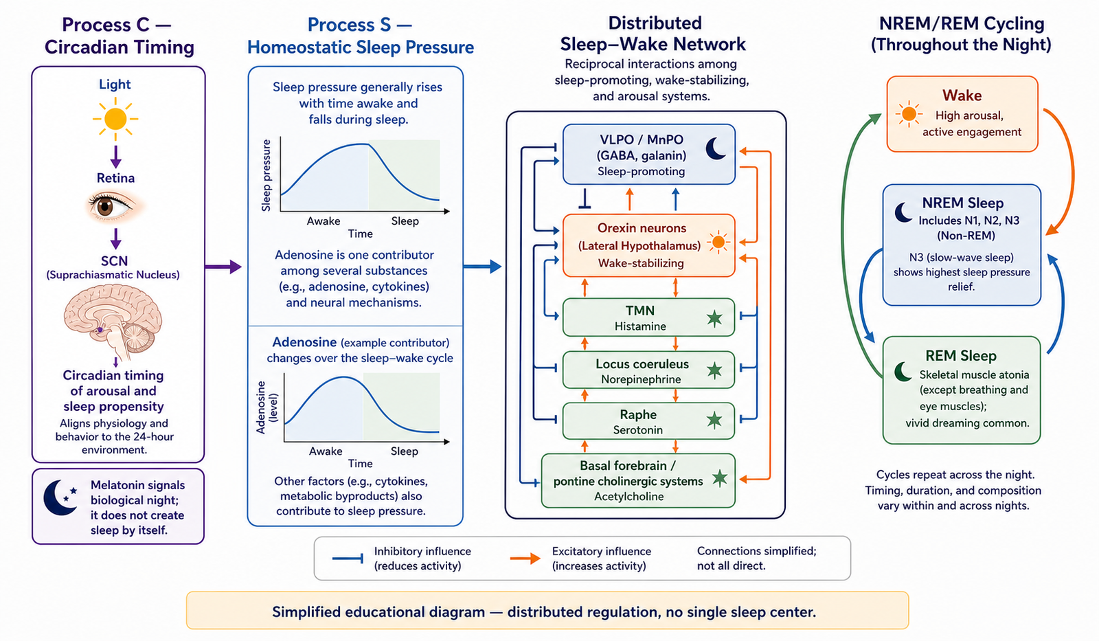
Sleep science conventionally separates sleep control into two interacting processes (the "two-process model," Borbély 1982; reaffirmed and refined in later work):
- Process C (circadian): A roughly 24-hour rhythm generated mainly by the SCN that sets the timing of the sleep–wake cycle and body functions (temperature, hormone release) relative to external light–dark cues.
- Process S (homeostatic sleep pressure): A drive that rises the longer a person is awake and falls during sleep, thought to reflect, among other things, accumulation and clearance of adenosine and other sleep-promoting substances in the brain.
A useful — but imperfect — analogy: Process C is like a timetable that says when the "sleep train" is scheduled to depart, while Process S is like a fuel gauge that says how urgently the body needs that trip. Neither factor alone explains sleep timing; they interact continuously. (Limitation of the analogy: real sleep regulation involves nonlinear interactions and additional oscillators, not a simple additive schedule.)
Within sleep itself, the brain further alternates between two qualitatively different states:
- NREM (non-rapid eye movement) sleep, staged as N1, N2, N3 under current American Academy of Sleep Medicine (AASM) criteria (N3 = slow-wave/"deep" sleep).
- REM (rapid eye movement) sleep, associated with vivid dreaming, muscle atonia, and fast, low-amplitude EEG activity resembling wakefulness.
2. Related Anatomy, Physiology, and Mechanisms
Sleep–wake control is distributed across several interacting brain regions rather than located in one "sleep center." Key structures include:
- Suprachiasmatic nucleus (SCN), hypothalamus: The brain's primary circadian pacemaker. It receives direct light input from specialized retinal ganglion cells (containing melanopsin) via the retinohypothalamic tract, allowing daily light exposure to reset ("entrain") the internal clock. The SCN does not itself produce sleep; it times when sleep-promoting and wake-promoting systems are favored.
- Ventrolateral preoptic nucleus (VLPO), hypothalamus: Contains GABA- and galanin-releasing neurons that are most active during sleep and inhibit wake-promoting arousal centers.
- Wake-promoting arousal systems: Include histamine neurons of the tuberomammillary nucleus, norepinephrine neurons of the locus coeruleus, serotonin neurons of the raphe nuclei, acetylcholine neurons of the basal forebrain and brainstem, and orexin/hypocretin neurons of the lateral hypothalamus, which help stabilize wakefulness.
- Basal forebrain: Contributes to both arousal regulation and to adenosine-mediated homeostatic sleep pressure.
- Brainstem structures (pons/midbrain, e.g., pedunculopontine and laterodorsal tegmental nuclei): Central to generating REM sleep features, including muscle atonia and rapid eye movements.
- Pineal gland: Produces melatonin under SCN control via a sympathetic pathway (SCN → paraventricular nucleus → spinal cord → superior cervical ganglion → pineal gland).
The "flip-flop switch" model
Sleep researchers (notably Saper and colleagues) proposed that sleep-promoting (VLPO) and wake-promoting (monoaminergic/orexin) neuron groups mutually inhibit one another, similar to an electrical flip-flop switch: when one side is active, it suppresses the other, producing relatively rapid, stable transitions between wake and sleep rather than a gradual blend. Orexin neurons are thought to help stabilize this switch, which is one reason their loss (as in narcolepsy) is associated with unstable, fragmented transitions between wakefulness, NREM, and REM sleep.
Homeostatic sleep pressure and adenosine
During wakefulness, adenosine — a byproduct of cellular energy use — is thought to accumulate in parts of the basal forebrain and cortex and to promote sleepiness partly by inhibiting wake-promoting neurons. This is one widely cited (but not the only proposed) mechanism underlying Process S. Caffeine's stimulant effect is generally attributed to blocking adenosine receptors, temporarily masking (not eliminating) accumulated sleep pressure.
Melatonin: a timing signal, not a sedative "sleep switch"
Light information from the retina reaches the SCN, which — in darkness — permits sympathetic signaling to the pineal gland, triggering melatonin synthesis and release. Melatonin levels normally rise in the evening and fall toward morning. Melatonin helps signal biological night to the rest of the body (a "timing" or synchronizing hormone) and can shift the timing of the circadian clock; it is not the primary substance that "produces" sleep itself, and evidence for its sedative effect size in otherwise healthy people is more modest and more variable than popular framing often suggests. Exogenous melatonin is used clinically mainly for circadian-timing problems (e.g., jet lag, some shift-work and delayed sleep phase situations) rather than as a general hypnotic, and regulatory status varies by country.
Glymphatic clearance — an emerging, not fully settled, area
A body of animal research (notably Xie et al., 2013, in mice) reported that clearance of interstitial waste products through the brain's perivascular ("glymphatic") pathways increases during sleep compared with wakefulness, and later human imaging/biomarker studies have reported findings consistent with sleep-associated brain clearance. However, this remains an area of active scientific debate: more recent tracer studies (2024–2025) have challenged aspects of the original mouse methodology and questioned how directly the animal findings translate to humans. MedMaru does not present glymphatic clearance as a proven, complete explanation of why sleep is restorative, nor as a basis for any treatment claim. It should be described as a promising but still-debated hypothesis. [Status of the controversy should be re-checked at time of publication, since this is a fast-moving research area.]
3. Step-by-Step: How a Typical Night Unfolds
- Evening — circadian "gate" opens. As external light decreases, the SCN reduces its inhibition on melatonin release. Homeostatic sleep pressure (Process S), which has been rising since morning wake time, is now high. The two processes align to increase sleep propensity.
- Sleep onset. VLPO neuron activity increases, inhibiting wake-promoting arousal centers (histamine, norepinephrine, serotonin, orexin systems). This is the flip-flop switch moving toward the "sleep" state.
- NREM progression (N1 → N2 → N3). EEG activity slows from mixed-frequency wake patterns into theta activity (N1), then sleep spindles and K-complexes (N2), then high-amplitude slow waves (N3, the deepest NREM stage, concentrated in the first part of the night).
- Transition to REM sleep. Brainstem circuits (pons) become more active; EEG activity resembles wakefulness ("paradoxical sleep"), rapid eye movements occur, and descending brainstem/spinal pathways produce near-total skeletal muscle atonia (a protective mechanism, generally understood to reduce the risk of acting out dream content).
- Cycling. NREM and REM alternate in cycles roughly every 90–110 minutes across the night; N3 (deep sleep) predominates earlier in the night, while REM periods generally lengthen toward the morning as circadian and homeostatic influences shift.
- Morning — circadian "gate" closes. As light exposure increases and Process S has been discharged by preceding sleep, the SCN increasingly favors arousal-system activity, melatonin falls, and the flip-flop switch shifts back toward stable wakefulness.
(Exact cycle counts and durations vary by individual, age, prior sleep debt, and health status; the figures above describe general population tendencies, not fixed rules for any one person.)
4. NREM vs. REM Sleep — Comparison Table
Note: functional roles of specific sleep stages (e.g., which stage is "more important" for which type of memory) remain an active research area with mixed findings across studies; the table reflects commonly cited associations, not settled consensus.
5. Clinical Relevance and Limits of Current Understanding
- Circadian rhythm sleep–wake disorders (e.g., delayed sleep phase, jet lag, shift-work disorder) reflect a mismatch between the SCN-driven timing system and the desired or required sleep schedule, not simply "poor sleep habits."
- Narcolepsy type 1 is associated with loss of orexin/hypocretin-producing neurons and destabilization of the wake–REM–NREM switching system, which is one explanation offered for symptoms such as excessive daytime sleepiness, cataplexy, and abnormal REM intrusion into wakefulness.
- Insomnia and other sleep disorders can involve disturbances of either the circadian process, the homeostatic process, or both, plus psychological and behavioral contributors — it is rarely explained by a single brain region or single neurotransmitter.
- Shift work, jet lag, and irregular light exposure can desynchronize the circadian and homeostatic processes from each other and from the external environment, which is one contributor (among several) to associated health effects reported in the research literature.
- Limitations: Much of the detailed cellular/circuit-level evidence (flip-flop switch mechanics, orexin stabilization, some adenosine mechanisms) comes primarily from animal models; the degree of direct translation to intact human physiology is an active area of research and should not be overstated as fully proven in humans. Sleep-stage "functions" (e.g., specific memory roles) remain hypotheses under ongoing investigation, not settled facts.
- This article is educational. It does not diagnose or treat any individual and is not a substitute for a clinical evaluation by a qualified healthcare professional (e.g., a sleep medicine specialist), especially for persistent insomnia, suspected sleep apnea, excessive daytime sleepiness, cataplexy, or parasomnias with injury risk.
6. Common Misconceptions vs. Accurate Explanation
7. Key Terms (for learners)
- Circadian rhythm: An internally generated, roughly 24-hour cycle in physiology and behavior, normally synchronized ("entrained") to the external light–dark cycle.
- Suprachiasmatic nucleus (SCN): A small hypothalamic structure that serves as the brain's main circadian pacemaker.
- Homeostatic sleep pressure (Process S): A drive to sleep that builds during wakefulness and dissipates during sleep.
- Two-process model: The framework (Borbély) describing sleep timing as the interaction of circadian (Process C) and homeostatic (Process S) influences.
- Flip-flop switch: A model describing mutual inhibition between sleep-promoting and wake-promoting neuron groups that produces relatively sharp transitions between states.
- VLPO (ventrolateral preoptic nucleus): A hypothalamic region containing sleep-promoting neurons.
- Orexin/hypocretin: Neuropeptides from the lateral hypothalamus that help stabilize wakefulness; their loss is linked to narcolepsy type 1.
- Adenosine: A molecule that accumulates during wakefulness and is thought to contribute to sleep pressure; blocked by caffeine.
- Melatonin: A pineal hormone released mainly at night under SCN control; functions as a circadian timing signal.
- NREM sleep (N1–N3): Non-rapid eye movement sleep stages, including slow-wave ("deep") sleep in N3.
- REM sleep: Rapid eye movement sleep, associated with vivid dreaming and near-total muscle atonia.
- Glymphatic system: A perivascular fluid pathway proposed to assist waste clearance in the brain, studied mainly in animal models with emerging, debated human evidence.
8. FAQ
Q1. Is there one single "sleep center" in the brain?
No. Sleep is generated by a distributed network — the SCN provides timing, the VLPO and arousal nuclei drive state switching, and brainstem circuits generate REM-specific features. No single structure alone "creates" sleep.
Q2. Does melatonin make you fall asleep the way a sedative does?
Not exactly. Melatonin mainly signals that it is biological night and helps align the timing of sleep with the circadian clock. Its direct sedative effect in healthy adults tends to be modest compared with popular claims, and its main evidence-based clinical uses relate to circadian timing problems rather than general sleeplessness.
Q3. Why do we need both NREM and REM sleep?
NREM (especially N3/slow-wave sleep) is associated with physical restoration and aspects of memory processing, while REM is associated with vivid dreaming and other proposed roles in emotional and procedural memory processing. Exact functional contributions of each stage remain an active research question, not a fully settled answer.
Q4. Does the brain "clean itself" only during sleep?
Animal research (mainly in mice) suggests waste clearance through perivascular ("glymphatic") pathways may be more active during sleep than wakefulness, and some human studies report findings consistent with this. However, this remains a debated and evolving research area, and it should not be presented as a settled or complete explanation for why sleep is needed, or as evidence for any specific treatment.
Q5. What controls when I feel sleepy versus wide awake during the day?
The interaction of your circadian clock (mainly SCN-driven) and homeostatic sleep pressure (rising with time awake) — plus factors such as light exposure, prior sleep debt, caffeine, and individual variation — together shape daytime alertness and sleepiness.
Q6. Can jet lag or shift work "break" this system?
They can desynchronize the circadian clock from the external light–dark cycle and from the homeostatic process, contributing to disrupted sleep timing and quality. This is generally reversible with time and light exposure management for most people, though persistent circadian sleep–wake disorders may need clinical evaluation.
9. Key Takeaways
- Sleep is actively generated and regulated by an interacting brain network, not a passive shutdown state.
- Two processes — a circadian clock (mainly the SCN) and homeostatic sleep pressure (linked to adenosine and time awake) — together determine sleep timing and intensity (two-process model, Borbély).
- A "flip-flop switch" of mutually inhibiting sleep- and wake-promoting neuron groups (VLPO vs. arousal/orexin systems) produces relatively sharp transitions between wake, NREM, and REM sleep.
- Melatonin is best understood as a circadian timing signal from the pineal gland, not a direct sleep-inducing sedative.
- NREM and REM sleep are physiologically distinct states with different EEG patterns, muscle tone, and proposed (not fully settled) functions.
- Glymphatic brain clearance during sleep is a promising but still scientifically debated hypothesis, based mainly on animal studies, and should not be presented as a proven treatment mechanism.
- Much circuit-level detail is drawn from animal research; translation to intact human physiology is still being actively studied.
10. Educational Purpose Statement and When to Seek Care
This article is for general education about brain physiology and is not personal medical advice, a diagnosis, or a treatment recommendation. Normal variation in sleep needs and patterns is common. However, consider seeking evaluation from a qualified healthcare professional (e.g., a primary care physician or sleep medicine specialist) if you or someone you know experiences:
- Ongoing difficulty falling or staying asleep that affects daily functioning
- Loud snoring with witnessed pauses in breathing, gasping, or excessive daytime sleepiness (possible signs of sleep apnea)
- Sudden muscle weakness triggered by strong emotion, or overwhelming daytime sleepiness (possible signs warranting narcolepsy evaluation)
- Acting out dreams physically, injury during sleep, or unsafe sleepwalking behavior
- Significant, persistent changes in sleep pattern with no clear cause
This is educational information only, not an emergency resource. If someone is in acute medical distress, contact local emergency services.
Related Articles
References
- Borbély AA. A two process model of sleep regulation. Human Neurobiology. 1982;1(3):195–204. https://pubmed.ncbi.nlm.nih.gov/7185792/
- Saper CB, Chou TC, Scammell TE. The sleep switch: hypothalamic control of sleep and wakefulness. Trends in Neurosciences. 2001;24(12):726–731. https://doi.org/10.1016/S0166-2236(00)02002-6
- Scammell TE, Arrigoni E, Lipton JO. Neural circuitry of wakefulness and sleep. Neuron. 2017;93(4):747–765. https://doi.org/10.1016/j.neuron.2017.01.014
- Porkka-Heiskanen T, Strecker RE, Thakkar M, et al. Adenosine: a mediator of the sleep-inducing effects of prolonged wakefulness. Science. 1997;276(5316):1265–1268. https://doi.org/10.1126/science.276.5316.1265
- Xie L, Kang H, Xu Q, et al. Sleep drives metabolite clearance from the adult brain. Science. 2013;342(6156):373–377. https://doi.org/10.1126/science.1241224
Written by: MedMaru Editorial Team
Reviewed for medical accuracy by:
S. Chang, KMD