What Does the Hippocampus Do? Memory, Location, and Anatomy
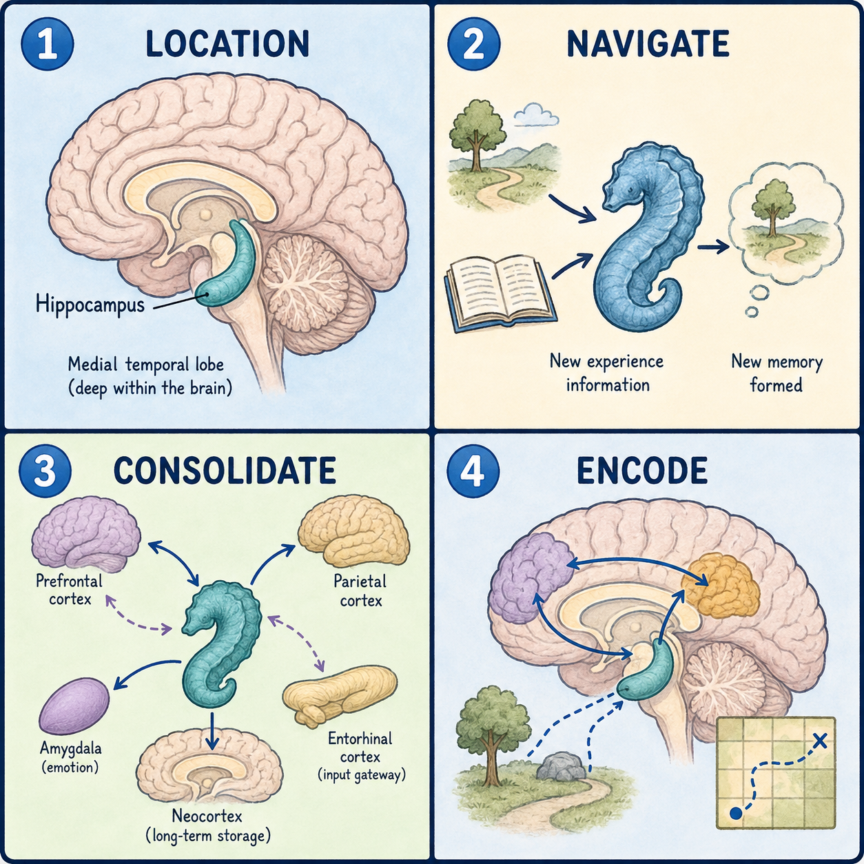
The hippocampus turns new experiences into memories that last. It is a curved, seahorse-shaped structure buried deep in the temporal lobe of the brain, and most people have two of them, one on each side. Together they are essential for forming new long-term memories and for finding your way around physical space.
The clearest way to see what the hippocampus does is to look at what happens without it. Someone who loses both hippocampi can still hold a thought in mind for a few seconds. They can still recall memories formed years earlier. What they cannot do is lay down new memories that last. That pattern was first documented in detail in a single, now-famous patient, and much of what follows grows out of his story.
Why it is called the hippocampus
The name comes from the Greek word for seahorse. Early anatomists thought the structure looked like one when they cut into the brain and saw it in cross-section. The comparison is fair. The hippocampus curves along the floor of the temporal horn of the lateral ventricle and tapers at both ends.
Under a microscope, it is not one uniform sheet of tissue. It is built from a few tightly interlocking parts that neuroscientists group together as the hippocampal formation: the dentate gyrus, the Cornu Ammonis fields (commonly labeled CA1 through CA4), and the subiculum. Signals generally flow in one direction through this circuit. They arrive mainly from the entorhinal cortex, pass through the dentate gyrus and then the CA fields, and leave through the subiculum. This one-way loop is thought to be central to how the brain turns a fleeting experience into a stored memory.
Where is the hippocampus located?
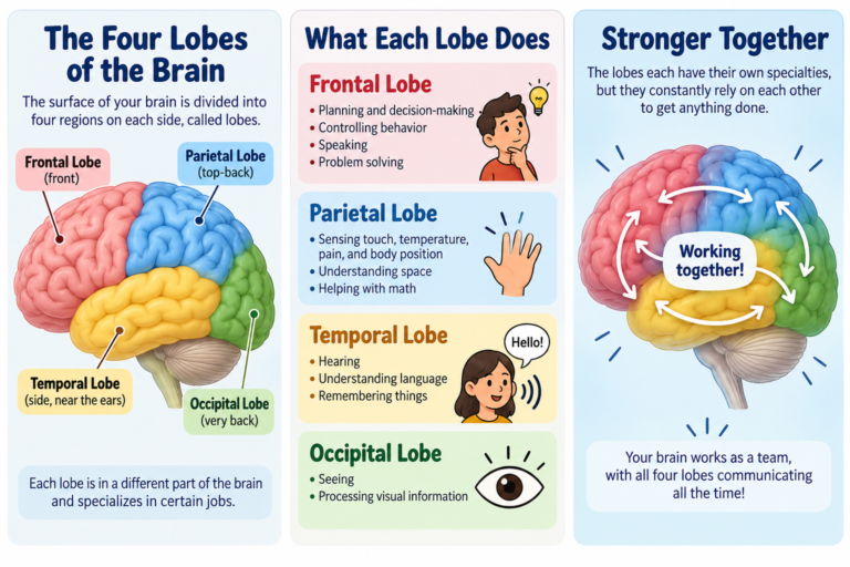
Each hippocampus sits within the medial temporal lobe, the inner surface of the temporal lobe, curled next to a few structures it works closely with. The amygdala sits just in front of it and adds emotional weight to memories. The entorhinal cortex acts as the main gateway, funneling information in from the rest of the cerebral cortex and back out again. A curved bundle of fibers called the fornix carries output from the hippocampus toward the hypothalamus and other targets involved in memory and in regulating the body’s internal state.
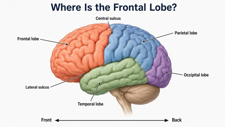
Because the hippocampus is tucked inside the temporal lobe rather than lying on the brain’s surface, you cannot see it from the outside. It only comes into view when the temporal lobe is dissected or imaged with MRI.
What does the hippocampus do in memory?
The hippocampus’s best-established job is converting short-term experience into long-term, consciously recallable memory. This is called declarative, or explicit, memory. It covers two related kinds: episodic memory, for specific personal events such as what you had for breakfast, and semantic memory, for general facts such as Paris being the capital of France.
A common picture is that the hippocampus is a filing cabinet where memories are kept for good. The leading model says otherwise. The hippocampus acts more like an index or a temporary assembly site. It binds together the scattered pieces of an experience, such as sights, sounds, and context, which are otherwise processed in separate regions of the cortex. Over time, through a process that unfolds during sleep and rest, those memories are gradually transferred to the cortex for long-term storage. This is why hippocampal damage tends to spare old memories formed long before the injury, even while it wipes out the ability to form new ones.
The two hippocampi also work together rather than splitting memory neatly in half. Some studies suggest a modest division of labor, with verbal memory depending more on the left side and spatial memory more on the right, but this division is not absolute.
How the hippocampus helps you find your way
The hippocampus also plays a central role in spatial memory and navigation. Certain hippocampal neurons, called place cells, fire selectively when an animal or person is in a specific location. Together they build an internal map of the surrounding environment, which is what lets you remember where things are and how to get from one place to another.
What happens when the hippocampus is damaged?
The most influential evidence comes from a patient known for decades in the scientific literature by his initials, H.M. In 1953 he underwent surgery to control severe epilepsy that did not respond to medication. The operation removed the medial temporal lobes on both sides, including roughly two-thirds of the hippocampal complex on each side. His seizures improved, but he was left with dense anterograde amnesia: he could no longer form new long-term memories of events or facts, yet his memories from before the surgery, his intelligence, and his ability to hold information for a few seconds all remained intact.
His case, reported by Scoville and Milner, became one of the foundational pieces of evidence that the hippocampus has a specific role in memory rather than being one interchangeable part of a general “memory organ.” It also answers a question people often ask: can you live without a hippocampus? You can. H.M. lived on after the operation with his personality, intelligence, and old memories preserved, while new long-term declarative memories no longer formed.
Beyond surgical cases, the hippocampus is one of the structures most often involved in two common neurological conditions. In temporal lobe epilepsy, part of the hippocampus frequently shows a pattern of scarring and neuron loss called hippocampal sclerosis, which is associated with seizures that start in that region. In Alzheimer’s disease, the hippocampus, and particularly the CA1 subregion, is typically among the earliest areas to lose cells. This is part of why early memory difficulty is such a prominent symptom of the disease.
Whether hippocampal damage is permanent depends on the cause and on how much tissue is affected. Some causes, such as prolonged oxygen deprivation or surgical removal, tend to produce lasting memory problems. Others may allow partial recovery, depending on the extent of the damage and the underlying condition.
This article describes normal anatomy and function for learning purposes. It is not a diagnosis and does not replace assessment by a clinician.
References
- Rao, N.P., Alsayouri, K. “Neuroanatomy, Hippocampus.” StatPearls. NCBI Bookshelf — https://www.ncbi.nlm.nih.gov/books/NBK482171/
- Scoville, W.B., Milner, B. “Loss of recent memory after bilateral hippocampal lesions.” Journal of Neurology, Neurosurgery, and Psychiatry, 1957;20(1):11-21.
- Voss, J.L., Bridge, D.J., Cohen, N.J., Walker, J.A. “A closer look at the hippocampus and memory.” Trends in Cognitive Sciences, 2017 — https://pmc.ncbi.nlm.nih.gov/articles/PMC5659202/
- Kenhub. “Hippocampus: Anatomy, functions and connections.” — https://www.kenhub.com/en/library/anatomy/hippocampus-structure-and-functions
Written by: MedMaru Editorial Team
Reviewed for medical accuracy by:
S. Chang, KMD