Internal Capsule: Location, Function, and Anatomy
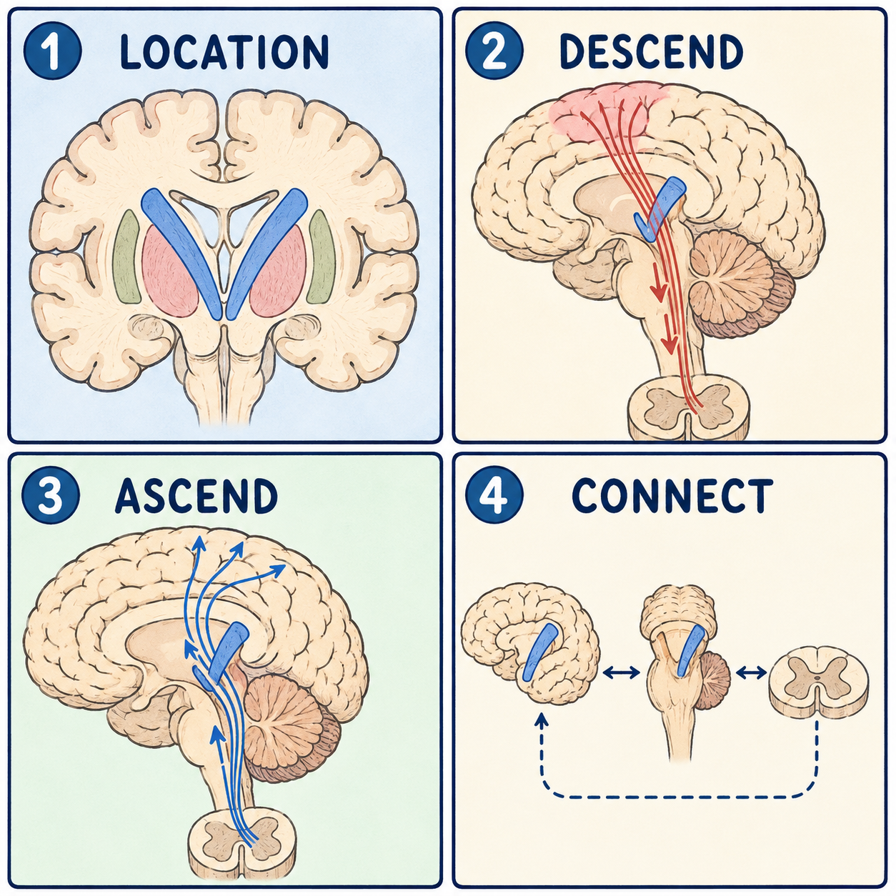
The internal capsule is a compact band of white matter buried deep inside each half of the brain. White matter is tissue made mostly of nerve fibers coated in myelin, a fatty insulation that speeds up signals. Almost every message traveling between the cerebral cortex and the deeper parts of the nervous system has to pass through this one narrow lane. That is why even a very small area of damage here, often from a stroke, can cause surprisingly large and specific problems.
A narrow corridor squeezed between gray matter
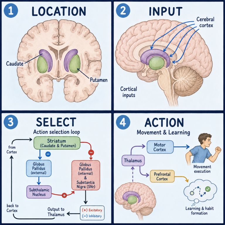
The internal capsule lies in the inner part of each cerebral hemisphere. On its inner side are the thalamus and the caudate nucleus. On its outer side are the putamen and globus pallidus, which together are called the lentiform nucleus because of their lens-like shape. It is this sandwiching between gray-matter structures that earned it the name capsule. A common mistake is to assume it is gray matter like its neighbors. It isn’t. It is a sheet of fibers wedged between them, carrying signals in both directions between the cortex and the thalamus, brainstem, and spinal cord.
Picture a highway interchange squeezed between two office buildings, with the thalamus on one side and the lentiform nucleus on the other. Many lanes of traffic, for movement, touch, vision, and hearing, are forced through one tight corridor. The analogy breaks down in one useful way. A real interchange has barriers between lanes, while these fiber tracts run side by side with no walls between them. That is part of why a single small lesion can affect several functions at once.
The internal capsule has two lesser-known relatives. Just outside the lentiform nucleus lies the external capsule, a thinner sheet of white matter separating the putamen from the claustrum. Farther out, between the claustrum and the insular cortex, lies the extreme capsule. Both carry association fibers that link one area of cortex to another, rather than the long pathways between cortex and brainstem or spinal cord that define the internal capsule. This article does not cover them as separate structures.
Five segments, each with its own traffic
Anatomists divide the internal capsule into five segments, and each one carries a different kind of traffic. The anterior limb is the front section. It carries fibers linking the thalamus with the frontal lobe, along with frontopontine fibers involved in planning and emotion-related processing. The genu, Latin for knee, is the bend that joins the front and back sections. It carries the corticobulbar tract, which controls the muscles of the face, jaw, and throat.
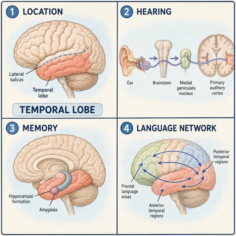
The posterior limb is the longest and busiest section. It carries the corticospinal tract, the main pathway for voluntary movement of the arms and legs, along with sensory fibers that relay touch and position sense. So the internal capsule is far from a purely motor structure. The retrolenticular part, just behind the lentiform nucleus, carries the optic radiation toward the visual cortex. The sublenticular part, just beneath it, carries the auditory radiation toward the temporal lobe.
Why it is a classic stroke site
Nerve cells across large areas of the cortex send fibers downward. As they approach the deep brain, those fibers funnel together and compress into the internal capsule’s five segments, sorted roughly by function. Most corticospinal fibers cross to the opposite side farther down. That is why damage on one side of the internal capsule usually causes weakness on the opposite side of the body.
The blood supply makes the area vulnerable. Small perforating arteries branching from the anterior cerebral, middle cerebral, anterior choroidal, and internal carotid arteries feed this compact, high-traffic zone. When one of these tiny arteries is blocked, the result is a lacunar stroke, a small, deep stroke named after the tiny cavity, or lake, that it leaves behind.
What damage looks like
Because so many pathways are packed into so little space, a lacunar stroke here tends to produce one of several recognizable syndromes, and which one depends on the segment involved. Damage to the posterior limb causes a pure motor stroke. The face, arm, and leg on one side become weak to a roughly equal degree, without any loss of sensation. When motor fibers and fibers involved in coordination are both affected, the result is ataxic hemiparesis, which combines weakness with clumsy, poorly coordinated movement. Damage near the anterior limb or genu causes clumsy-hand dysarthria, in which slurred speech comes paired with a clumsy hand. Larger hemorrhages can cause severe paralysis on the opposite side, along with spasticity and exaggerated reflexes.
These are recognizable patterns, not a perfect one-to-one map. The exact edge of a small lesion and normal variation in how fibers are arranged mean the same syndrome can arise from slightly different locations. Imaging is needed to confirm the cause.
This article is educational background, not a diagnostic guide. Sudden weakness on one side of the body, facial drooping, or slurred speech can signal a stroke and needs emergency medical care.
Related: What Is the Cerebral Cortex? · What Is the Brain? · What Is the Cerebrum?
References
- StatPearls. “Neuroanatomy, Internal Capsule.” NCBI Bookshelf, NBK542181 — https://www.ncbi.nlm.nih.gov/books/NBK542181/
Written by: MedMaru Editorial Team
Reviewed for medical accuracy by:
S. Chang, KMD