What Is the Parasympathetic Nervous System?
The parasympathetic nervous system handles the body’s routine maintenance. It digests food, slows the heart, produces saliva and tears, empties the bladder, and adjusts the eyes for close-up vision, all without you having to think about it. It is one of the divisions of the autonomic nervous system, the part of the nervous system that runs involuntary functions. Its fibers start in the brainstem and in the sacral part of the spinal cord, the vagus nerve carries its signals to the heart, lungs, and much of the gut, and it uses acetylcholine as its main chemical messenger. It is often called the rest-and-digest system. That phrase is useful but incomplete, because this system stays active all day and works continuously alongside the sympathetic nervous system to keep the body in balance.
The everyday work it does
Parasympathetic signals regulate internal organs without conscious effort, and they matter most during ordinary, non-emergency conditions. They slow the heart rate, constrict the pupils, help the eyes focus on nearby objects, stimulate tears and saliva, promote digestive secretions, speed the movement of food through the gut, assist urination and bowel movements, and contribute to sexual arousal. Together, these actions help the body process nutrients, get rid of waste, conserve energy, and keep internal conditions stable.
Two starting points: brainstem and sacrum
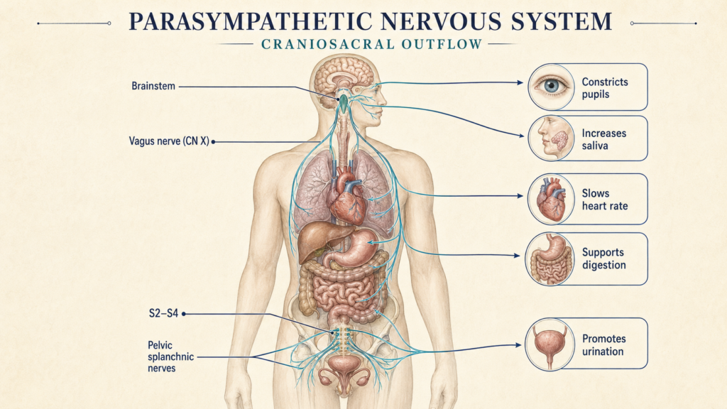
Parasympathetic signals come from two main regions, the brainstem and the sacral spinal cord. That is why the system is traditionally called the craniosacral division of the autonomic nervous system.
From the brainstem, parasympathetic fibers leave through four cranial nerves, each with its own territory. The oculomotor nerve, cranial nerve III, constricts the pupil and helps the eye focus on near objects. The facial nerve, cranial nerve VII, stimulates the tear glands and some salivary glands. The glossopharyngeal nerve, cranial nerve IX, stimulates the parotid salivary gland. The vagus nerve, cranial nerve X, supplies the heart, lungs, and much of the digestive tract. Of the four, the vagus has by far the widest reach, carrying signals from the brainstem down through the neck and chest into the abdomen.
From the sacral spinal cord, parasympathetic fibers arise from segments S2 to S4. They travel through the pelvic splanchnic nerves to the lower part of the large intestine, the rectum, the bladder, and parts of the reproductive system. This sacral route is especially important for urination, defecation, and sexual function.
A long wire and a short wire
A typical parasympathetic pathway uses two neurons in sequence. The first, the preganglionic neuron, starts in the brainstem or sacral spinal cord and sends its axon all the way to a small relay station, an autonomic ganglion, that sits close to or even inside the target organ. The second, the postganglionic neuron, starts in that ganglion and travels only a short distance to the organ.
Think of it like a long-distance delivery that hands the parcel to a local courier at the last stop. Because the relay sits near the organ, parasympathetic pathways generally have long preganglionic fibers and short postganglionic fibers. The sympathetic system is organized the other way around, with its relays usually closer to the spinal cord. The delivery picture is a simplification, since one preganglionic fiber may contact several postganglionic neurons, but it captures the layout.
The parasympathetic system uses acetylcholine at both steps. At the ganglion, acetylcholine from the preganglionic neuron binds nicotinic acetylcholine receptors. At the target organ, acetylcholine from the postganglionic neuron usually binds muscarinic receptors. Different muscarinic subtypes produce different effects. M2 receptors in the heart help slow the heart rate, while M3 receptors contribute to gland secretion and smooth-muscle contraction.
Organ by organ
In the eyes, parasympathetic activity constricts the pupils and supports near vision, and it increases tear production. It increases watery saliva, slows the heart, constricts the smooth muscle of the airways and increases their secretions, and boosts movement and secretion in the stomach and intestines. It supports pancreatic secretion during digestion and contributes to the neural control of gallbladder contraction. In the bladder, it contracts the detrusor muscle and helps the outlet relax, supporting urination. It also supports defecation and contributes to genital blood flow and erection. These are general patterns, and the actual response depends on the organ, the receptor type, and the situation.
The vagus nerve’s special role
The vagus nerve is the main parasympathetic link between the brainstem and the organs of the chest and abdomen. Its branches influence heart rate, airway tone, digestive secretions, and the movement of food through the gut. It is not purely a parasympathetic motor nerve, though. It also contains sensory fibers that carry information from internal organs back to the brain, helping the brain monitor the heart, lungs, and digestive system. And it doesn’t reach everything. Pelvic organs and the lower large intestine get their parasympathetic supply mainly from the sacral spinal cord.
Not simply the opposite of sympathetic
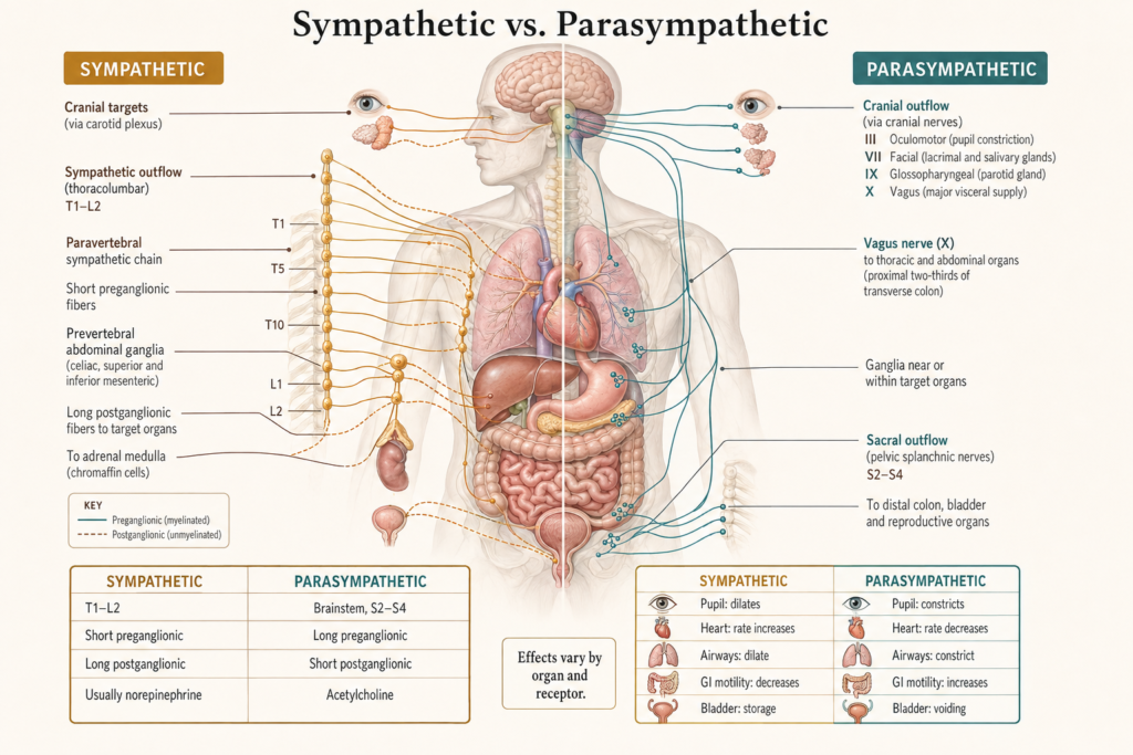
The parasympathetic and sympathetic systems sometimes pull in opposite directions, but they are not simple on-off switches. Sympathetic activity tends to speed the heart while parasympathetic activity slows it. Sympathetic activity widens the pupils while parasympathetic activity narrows them. During acute stress, sympathetic activity can slow the gut, while parasympathetic activity generally promotes digestion.
In other organs the relationship is more complicated. Some tissues are controlled mainly by one division, and some functions need both working together. Sweat glands, most blood vessels, and the tiny muscles that raise body hair are controlled mainly by sympathetic pathways rather than direct parasympathetic nerves.
Anatomically, the two divisions also differ. The parasympathetic system starts in the brainstem and S2 to S4 spinal cord, while the sympathetic system, the thoracolumbar division, starts in the thoracic and upper lumbar cord. The sympathetic system’s main postganglionic messenger is usually norepinephrine rather than acetylcholine, and its general role is mobilizing the body for physical or emotional demands. Both remain active and cooperate as conditions change.
Why rest and digest is only half the story
The rest-and-digest label can create two misunderstandings. First, the parasympathetic system doesn’t switch on only when you are resting. Its signals continuously contribute to heart-rate control, digestion, secretion, and other normal functions. Second, parasympathetic activity doesn’t mean the whole body is calm. Autonomic control is organized organ by organ, according to need, and parasympathetic activity may rise in one pathway while other autonomic responses are happening elsewhere. A more accurate description is that the parasympathetic system supports routine organ regulation, energy conservation, digestion, secretion, and elimination, working with sympathetic pathways and central autonomic circuits to maintain homeostasis.
When it doesn’t work properly
Abnormal parasympathetic signaling can affect several organs. Possible signs include an unusually fast or slow heart rate, dry eyes or mouth, trouble adjusting pupil size, impaired digestion, constipation, difficulty emptying the bladder, and sexual dysfunction. None of these is specific to parasympathetic problems. They can also come from medications, dehydration, endocrine disorders, nerve damage, or other conditions, and symptoms alone cannot show which part of the autonomic nervous system is affected.
This article is educational background, not a diagnostic guide. Persistent symptoms like these should be evaluated by a clinician.
Related: What Is the Autonomic Nervous System? · What Is the Sympathetic Nervous System? · What Is the Sympathetic Nervous System? · What Is the Nervous System? Structure and Main Functions
References
- Waxenbaum JA, Reddy V, Das JM. Anatomy, Autonomic Nervous System. StatPearls. Updated December 1, 2025. StatPearls Publishing. NCBI Bookshelf
- McCorry LK. Physiology of the autonomic nervous system. American Journal of Pharmaceutical Education. 2007;71(4):78. doi:10.5688/aj710478. PubMed Central
- Tindle J, Tadi P. Neuroanatomy, Parasympathetic Nervous System. StatPearls. Updated October 31, 2022. StatPearls Publishing.
- MSD Manual Professional Edition. Overview of the Autonomic Nervous System. Reviewed May 2025. MSD Manual
Written by: MedMaru Editorial Team
Reviewed for medical accuracy by:
S. Chang, KMD