Substantia Nigra: Location, Function, and Anatomy
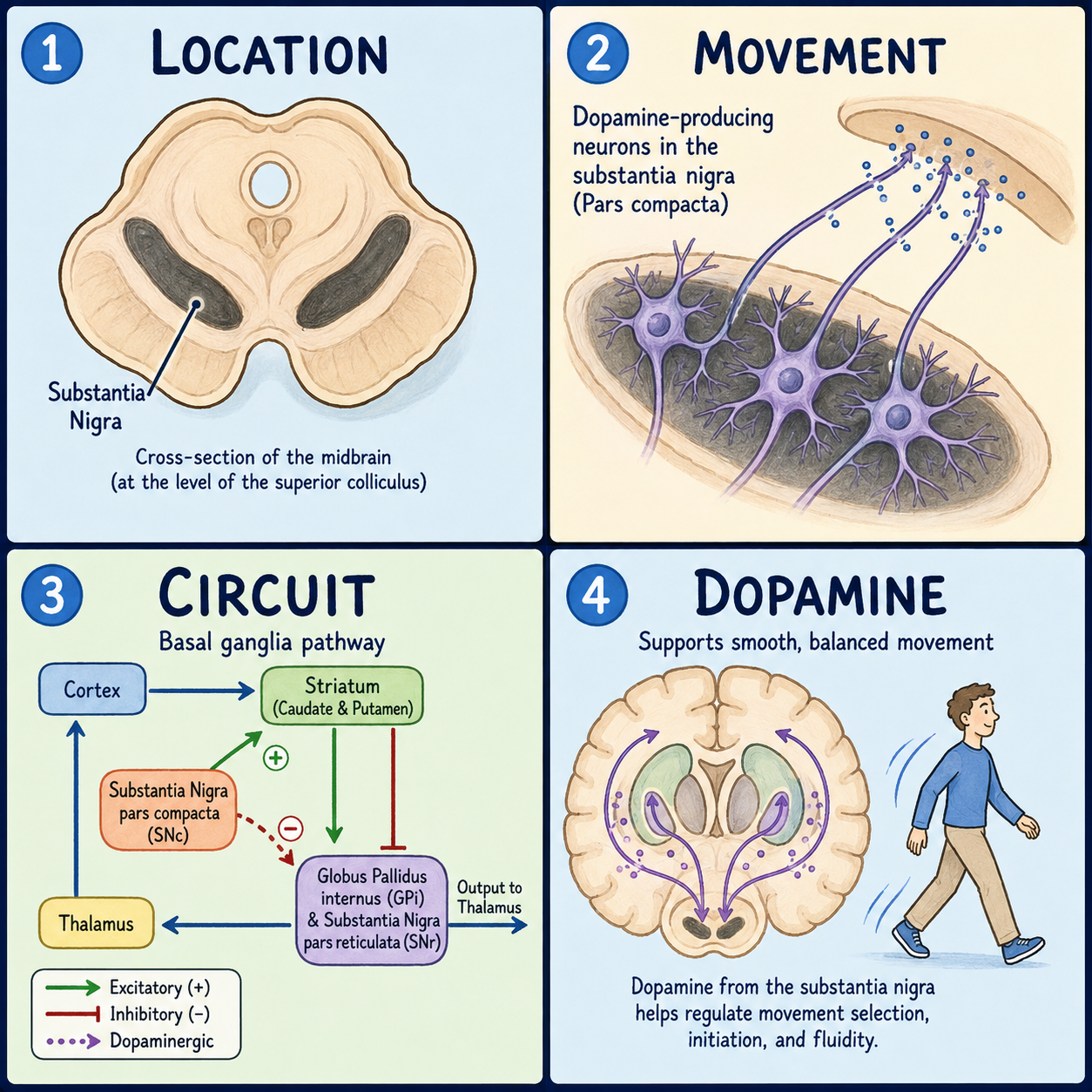
The substantia nigra is a small, dark strip of tissue buried deep in the midbrain, the upper part of the brainstem. Its best-known job is producing dopamine, the chemical messenger that helps the brain plan and smooth out movement. There are two of them, one on each side of the midbrain, because the brain has two halves. Each connects closely to a structure called the striatum through a pathway that fails in Parkinson’s disease.
Why it is black, and why it has two layers
“Substantia nigra” is Latin for “black substance,” which is literally what an anatomist sees when this tissue is cut: a dark band running through an otherwise pale slice of midbrain. The color comes from neuromelanin, a dark pigment that builds up inside certain neurons as they process dopamine over a lifetime.
It isn’t one uniform strip. It splits into two layers lying back to back. The pars compacta, closer to the middle of the brain, is made mostly of the dopamine-producing neurons that give the structure its color. The pars reticulata, closer to the surface, is made of different neurons that use GABA, an inhibitory messenger that tells other brain cells to slow down, instead of dopamine.
Think of it as two floors of the same small building. The upper floor manufactures dopamine and ships it to a distant warehouse, the striatum. The lower floor runs a separate switchboard, helping decide which movement signals get through. The floors aren’t simply stacked, though. They exchange signals with each other, so it isn’t a one-way delivery chain.
Where it sits
The substantia nigra lies inside the midbrain, just above where the brainstem narrows into the pons. It sits directly behind a fiber bundle called the crus cerebri, which carries the cortex’s outgoing movement commands down toward the spinal cord. That placement isn’t incidental, since sitting right where those signals pass lets it help fine-tune them. The pars compacta sits closer to another midbrain structure, the red nucleus. The pars reticulata sits closer to the crus cerebri and works alongside the globus pallidus internus, a basal ganglia structure located farther forward in the brain.
Although it is usually discussed together with the basal ganglia and wired tightly into their circuits, the substantia nigra is anatomically part of the midbrain, not the same forebrain region as the striatum and globus pallidus. It is also a separate structure from the striatum; the two are connected by the nigrostriatal pathway, not the same thing.
The pathway that fails in Parkinson’s
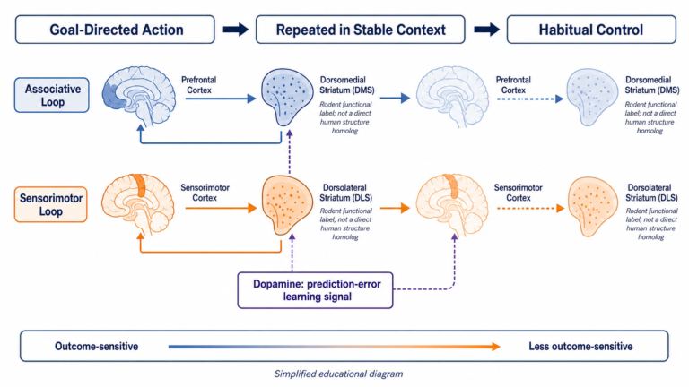
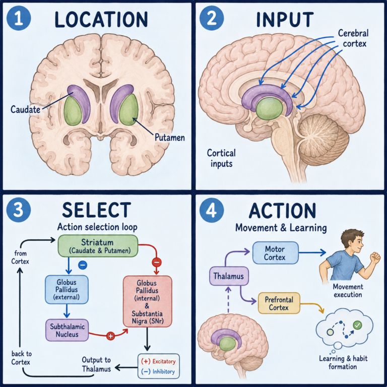
The nigrostriatal pathway carries dopamine from the pars compacta up to the striatum, the main entry point of the basal ganglia, the group of structures that helps select and smooth voluntary movement. Once dopamine arrives, it acts on two kinds of receiving neurons. One kind carries D1-type receptors that make movement easier to start. The other carries D2-type receptors that help hold unwanted movement back.
The pars reticulata, meanwhile, works with the globus pallidus internus as one of the basal ganglia’s main output routes. Together they send a constant, low-level “hold still” signal to the thalamus. Movement happens when other basal ganglia circuits briefly interrupt that signal at the right moment.
When dopamine-producing neurons in the pars compacta die, dopamine delivery to the striatum drops, the direct and indirect pathways fall out of balance, and the hold-still signal grows too strong. Movement becomes harder to start and slower to carry out. That shows up as rest tremor, stiffness, slowness of movement (bradykinesia), and balance problems.
When it is damaged
The pars compacta is the structure most consistently damaged in Parkinson’s disease. By the time symptoms appear, most people have already lost more than half of these dopamine-producing cells, commonly cited as 60 to 80 percent or more. That is part of why Parkinson’s is usually well underway by the time it is diagnosed, and why researchers are looking for earlier markers of nigral damage, though none are yet part of routine clinical diagnosis. Diagnosis during life is made clinically, from a person’s symptoms and a neurological examination, not by imaging the substantia nigra directly. Substantial loss of pars compacta neurons is the defining finding at autopsy.
It is important not to oversimplify why the cell loss happens. A common shortcut says Parkinson’s is simply a dopamine deficiency. Reduced dopamine explains the motor symptoms, but not why the neurons die in the first place. In most people with Parkinson’s, the underlying cause is unknown. Some cases are inherited and tied to specific genetic changes, and researchers think overall risk comes from a mix of genetic and environmental factors, including pesticide and pollutant exposure in some people. Substantia nigra cell loss and reduced dopamine are the confirmed core pathology. They explain what goes wrong, not why it starts.
Current treatments manage symptoms, for example by replacing dopamine, rather than restoring lost neurons. Whether the damage itself can be slowed or reversed is still being researched.
This article is educational background, not a diagnostic guide. New tremor, stiffness, slowed movement, frequent falls, or other unexplained neurological symptoms should be evaluated by a clinician rather than matched against this article.
Related: What Is the Cerebral Cortex? · What Is the Brain? · What Is the Brainstem?
References
- StatPearls, “Neuroanatomy, Substantia Nigra” — https://www.ncbi.nlm.nih.gov/books/NBK536995/
- StatPearls, “Parkinson Disease” — https://www.ncbi.nlm.nih.gov/books/NBK470193/
- NINDS, “Parkinson’s Disease” — https://www.ninds.nih.gov/health-information/disorders/parkinsons-disease
- Cleveland Clinic, “Substantia Nigra (SN)” — https://my.clevelandclinic.org/health/body/23010-substantia-nigra-sn
Written by: MedMaru Editorial Team
Reviewed for medical accuracy by:
S. Chang, KMD