Basal Ganglia: Function, Location, and Their Role in Parkinson’s Disease
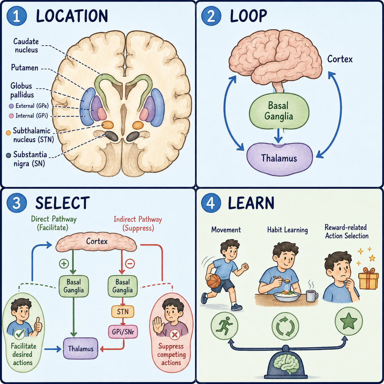
The basal ganglia are a group of deep brain structures that help decide which movements happen and how strongly. Think of them as gatekeepers sitting between your thinking brain and your moving muscles, deciding which movements get the green light and which get held back. In medical language, the basal ganglia, also called the basal nuclei, are a cluster of interconnected gray matter nuclei lying deep to the cerebral cortex. They have other roles too, but this article focuses on movement, because that is where the evidence is strongest and most consistent.
A team of nuclei, not a single organ
A common misconception treats the basal ganglia as one solid organ. They are actually a group of nuclei. A nucleus, in this sense, is a cluster of nerve cell bodies that work together. The members consistently included in the group are the caudate nucleus, the putamen, the globus pallidus, the subthalamic nucleus, and the substantia nigra. Two of them, the caudate and the putamen, are so closely linked in function that they are often described together as the striatum, the main entry point where signals from the cortex arrive.
A useful way to picture the whole team is a dimmer switch for movement rather than an on-off switch. A dimmer doesn’t simply turn a light fully on or fully off. It adjusts how bright the light is. In the same way, the basal ganglia mostly fine-tune how much a movement happens rather than starting it from nothing. The comparison has a limit: a real dimmer is one hand on one dial, while the basal ganglia run several looped circuits at once, so their dimming is the sum of many signals rather than one simple turn. This also corrects another myth. The basal ganglia don’t just start movement. They stay involved throughout an ongoing movement, filtering and adjusting which actions proceed and how vigorously.
Why they sit at a crossroads
The basal ganglia lie deep inside each cerebral hemisphere, beneath the cortex, and the position is not random. Signals for voluntary movement travel from the cortex down toward the spinal cord. The basal ganglia sit almost directly along that route, close to the thalamus, a relay station that passes signals back up to the cortex. That crossroads location lets them intercept and adjust movement commands before the commands are finalized, rather than reworking a movement after it has happened.
Each member has its own address. The caudate nucleus is C-shaped, with a head, body, and tail, and it sits lateral to the fluid-filled lateral ventricles. The putamen and the globus pallidus sit together just beneath the insula, and together they are called the lentiform nucleus. The lentiform nucleus is bounded on the inner side by the caudate and thalamus and on the outer side by a band of white matter called the external capsule.
The globus pallidus is itself split in two. The outer segment is the globus pallidus externus, or GPe, and the inner segment is the globus pallidus internus, or GPi. A thin sheet of white matter separates them. On its inner side, the globus pallidus is bordered by the curve of the internal capsule, a major white-matter highway that carries movement and sensory signals between the cortex and the spinal cord. That closeness matters clinically. Because the internal capsule and the basal ganglia touch, a stroke or bleed here can damage both at once, which is one reason basal ganglia strokes can cause movement problems.
The subthalamic nucleus sits just below the thalamus. The substantia nigra sits lower still, in the midbrain, and has two parts called the pars compacta and the pars reticulata. So most of the basal ganglia sit within the cerebral hemispheres, while the substantia nigra sits in the brainstem. It still counts as part of the group because its signals are wired so tightly into the same circuits.
Accelerator and brake: the direct and indirect pathways
The basal ganglia work as a loop. The cerebral cortex sends signals into the striatum. The striatum passes them through the globus pallidus and the substantia nigra. The output then returns to the cortex by way of the thalamus. Within this loop, the caudate and putamen act as input structures, while the GPi and the substantia nigra pars reticulata act as the main output structures that send signals to the thalamus. The pars compacta has a different job: it supplies dopamine to the striatum.
The loop runs through two main routes. In the direct pathway, striatal neurons send inhibitory, signal-dampening projections to the GPi and the substantia nigra pars reticulata. Quieting these output structures releases the thalamus from inhibition, so the thalamus can excite the motor cortex and a movement is allowed to go ahead. In the indirect pathway, striatal neurons inhibit the GPe instead. The GPe then reduces its own inhibition of the subthalamic nucleus. The now more active subthalamic nucleus excites the GPi and the pars reticulata, which increases inhibition of the thalamus and holds a movement back.
Think of a car with both an accelerator and a brake. The direct pathway acts something like the accelerator, helping a chosen movement go forward. The indirect pathway acts something like the brake, holding competing or unwanted movements in check. The limit here is that a car has one accelerator and one brake, while the basal ganglia run many of these loops in parallel, for many possible movements at once.
Because the circuit depends on a chain of connected structures, trouble almost anywhere along the loop can upset the balance between go and stop. When the direct pathway is favored too strongly, the result tends to be too much unwanted movement. When the indirect pathway is favored too strongly, the result tends to be too little movement. That is why basal ganglia damage does not always cause too little movement. It can cause either extreme, depending on which part of the circuit is affected.
It is also worth noting that the basal ganglia never control muscles directly. They adjust signals that pass through the thalamus back to the motor cortex, and it is the motor cortex that sends commands down to the spinal cord and muscles.
What Parkinson’s, Huntington’s, and hemiballism reveal
Most people first hear about the basal ganglia through Parkinson’s disease. In Parkinson’s, the dopamine-producing neurons of the substantia nigra pars compacta gradually degenerate. Dopamine normally helps tip the balance toward the direct, go pathway. When dopamine falls, the indirect, stop pathway becomes relatively stronger. That shift is thought to contribute to slowness of movement, called bradykinesia, along with resting tremor and a shuffling gait. This is a simplified circuit-level summary, not the whole story. Other brain regions and non-motor symptoms are involved, and basal ganglia involvement alone is not a diagnosis. Diagnosing Parkinson’s depends on clinical evaluation by a qualified clinician.
Huntington’s disease shows the opposite pattern. It is an inherited condition caused by an expanded CAG repeat, and it progressively kills neurons in the caudate and putamen. The damage falls heavily on cells that feed the indirect, stop pathway, so the go pathway becomes relatively unopposed. That is thought to contribute to chorea, the excessive, involuntary movements seen early in the disease, alongside cognitive and psychiatric changes.
Hemiballism is less common. It causes sudden, forceful, flinging movements of one arm and leg. It is typically linked to a lesion of the subthalamic nucleus on the side opposite the affected limbs, which removes an important brake on the direct pathway.
Blood supply adds one more clinical thread. The lenticulostriate arteries, small branches of the middle cerebral artery, are the main blood supply to this region. They are especially prone to rupture in people with poorly controlled high blood pressure, which makes the putamen one of the most common sites of hypertensive brain hemorrhage. That is a structural and vascular fact, not a statement about any one person’s risk.
Finally, a word on the model itself. The direct and indirect pathway picture is a widely used teaching framework, and it captures the basic logic of go versus stop signaling well. Basal ganglia research is an active field, though, and this two-pathway picture is a simplification rather than a claim that these are the only signaling routes involved.
This article is educational background and does not replace medical evaluation. New tremor, stiffness, slowness of movement, or involuntary movements should be assessed by a clinician.
Related: What Is the Cerebral Cortex? · What Is the Brain? · What Is the Cerebrum?
References
- StatPearls. “Neuroanatomy, Basal Ganglia.” NCBI Bookshelf, NBK537141 — https://www.ncbi.nlm.nih.gov/books/NBK537141/
- StatPearls. “Neuroanatomy, Globus Pallidus.” NCBI Bookshelf, NBK557755 — https://www.ncbi.nlm.nih.gov/books/NBK557755/
Written by: MedMaru Editorial Team
Reviewed for medical accuracy by:
S. Chang, KMD