Cerebral Cortex: Layers, Areas, and Function
The cerebral cortex is the thin, folded layer of tissue covering the two cerebral hemispheres, the large outer part of the brain. It helps you interpret what you sense, guide voluntary movement, use language, form memories, direct attention, and make decisions. Its surface is folded into ridges called gyri and grooves called sulci. Most of the human cortex is a six-layered type called the neocortex, and different regions specialize in different kinds of work. Still, the cortex doesn’t do any of this alone. Most complex abilities depend on networks linking many cortical areas with deeper structures such as the thalamus, basal ganglia, hippocampus, cerebellum, and brainstem.
A thin sheet with enormous reach
The cortex is a continuous sheet of neural tissue covering the cerebrum. In most places it is only a few millimeters thick, yet it holds vast numbers of neurons and connections. It counts as gray matter because it contains neuronal cell bodies, dendrites and synapses, glial cells, blood vessels, and unmyelinated stretches of axons. So gray matter is not just cell bodies.
It is also not the whole brain, which is a common confusion. The cerebrum includes the cortex, the white matter beneath it, and deep gray-matter structures. Beyond the cerebrum, the brain also includes the diencephalon, brainstem, and cerebellum.
Where it sits and why it is folded
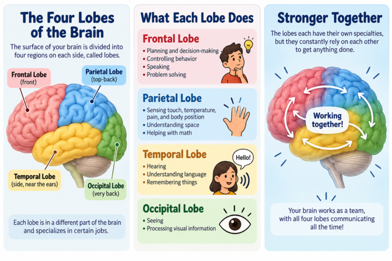
The cortex covers the outer surface of both hemispheres and dips down into the grooves between the folds. Beneath it lies cerebral white matter, packed with axons that connect cortical areas within the same hemisphere, link the two hemispheres, and tie the cortex to deeper structures, the brainstem, and the spinal cord. The cortex is traditionally divided into four lobes: frontal, parietal, temporal, and occipital. The insula, tucked deep inside the lateral sulcus, is sometimes described as a fifth.
Why all the folding? Think of crumpling a large sheet of paper to fit it into a small box. Folding lets a large cortical surface fit inside the skull. The analogy is loose, since the brain’s folds form in an orderly way during development rather than at random, but the space-saving idea holds. The folds also give anatomists landmarks. Particularly deep grooves are called fissures, and important ones include the central, lateral, parieto-occipital, and calcarine sulci. These landmarks help locate regions, but they don’t always mark exact functional boundaries.
Six layers, and the exceptions
The cortex can be classified by its microscopic organization. Most of the human cortex is neocortex, also called isocortex, which has six layers. Their thickness and cell makeup vary from region to region. Neocortex includes areas for body sensation, vision and hearing, voluntary movement, language, spatial processing, attention, and planning and decision-making. The name describes an anatomical and developmental type of cortex. It does not mean every neocortical region performs a higher cognitive function.
The six layers are numbered from the surface inward. Layer I, the molecular layer, has few cell bodies and many dendrites and horizontal axons. Layer II, the external granular layer, holds small neurons mainly involved in local processing. Layer III, the external pyramidal layer, holds pyramidal neurons that often connect with other cortical areas. Layer IV, the internal granular layer, receives prominent input from the thalamus and is especially well developed in primary sensory cortex. Layer V, the internal pyramidal layer, holds major output neurons that project to structures below the cortex, and it is particularly prominent in motor cortex. Layer VI, the multiform layer, contains several neuron types and sends important projections back to the thalamus. Because Layer IV dominates sensory areas and Layer V dominates motor areas, not every region looks the same under the microscope.
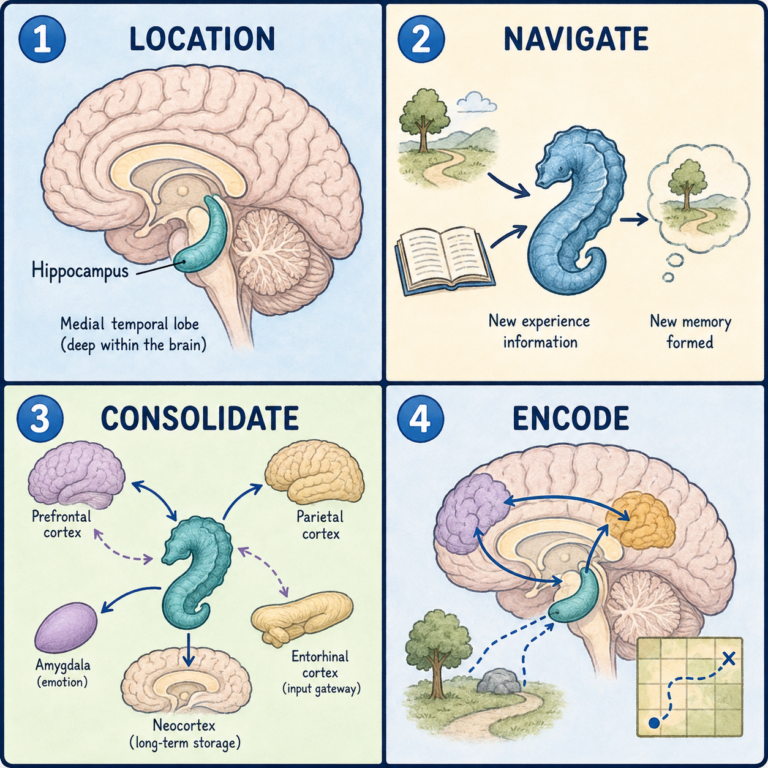
Not every part of the cortex has six layers, either. The allocortex has fewer than six clearly defined layers. Its main forms are the archicortex, closely tied to the hippocampal memory system, and the paleocortex, mainly associated with smell. There are also transitional regions between neocortex and allocortex, and the terminology varies between classification systems.
Sensory, motor, and association areas
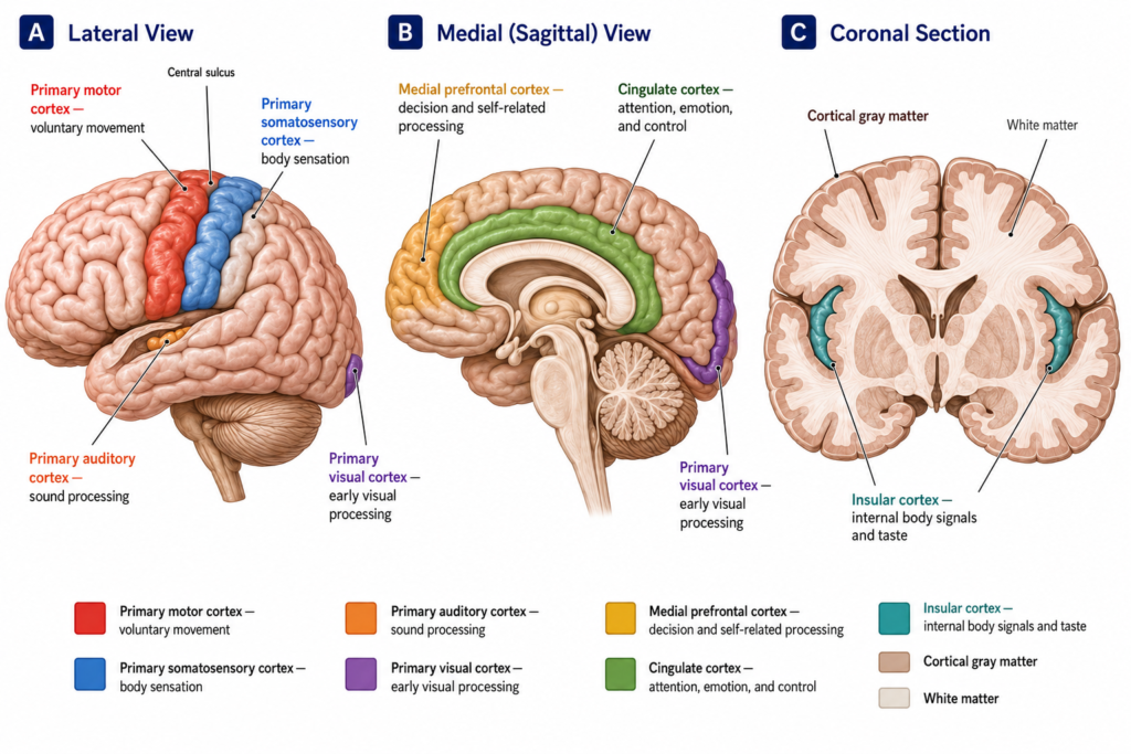
Cortical regions are often grouped into three kinds. Primary sensory areas receive major input from specific sensory pathways, usually by way of the thalamus. Examples are the primary somatosensory cortex in the parietal lobe, the primary visual cortex in the occipital lobe, and the primary auditory cortex in the temporal lobe. They process key features of sensory information, but complete perception depends on broader cortical and subcortical networks.
The primary motor cortex lies in the precentral gyrus of the frontal lobe and contributes to voluntary movement by sending signals down the descending motor pathways. Movement also depends on the premotor cortex, supplementary motor areas, sensory feedback, the basal ganglia, the cerebellum, the brainstem, and spinal circuits.
Association cortex is the broad term for areas outside the primary sensory and motor regions. These areas integrate information and contribute to recognizing objects and faces, language, spatial attention, working memory, planning, memory retrieval, and regulating behavior. They are essential parts of distributed networks, not independent centers for single abilities.
A network, not a set of control rooms
Cortical neurons form local circuits and also talk to distant regions through white-matter pathways. Signals pass between cortical areas and between the cortex and deeper structures. Visual signals, for instance, reach the primary visual cortex after traveling through subcortical pathways. The information is then processed across interconnected regions that analyze color, form, movement, identity, and location. Attention, memory, emotion, expectation, and current goals can all shape that processing.
That is why the cortex is better understood as a dynamic network than as a collection of independent control centers. Some functions do show clear anatomical specialization, but complex abilities such as language, memory, attention, and decision-making rely on distributed cortical and subcortical networks. The popular claim that humans use only 10 percent of the cortex has no scientific support. Activity shifts with the task and the person’s state, but normal brain function depends on widely distributed networks.
When the cortex is damaged
The cortex can be affected by stroke, traumatic brain injury, tumors, seizures, infections, inflammatory conditions, neurodegenerative diseases, and developmental abnormalities. Depending on the area, damage can cause weakness or impaired movement, reduced or altered sensation, loss of part of the visual field, difficulty speaking or understanding language, trouble recognizing objects or faces, impaired spatial awareness, memory or attention problems, changes in judgment or behavior, or seizures.
Symptoms depend on where the damage is, how extensive it is, which hemisphere is involved, and which connections are affected. Similar symptoms can also come from damage to white-matter pathways or subcortical structures, so a neurological examination and appropriate testing are needed to identify the affected area.
This article is educational background, not a diagnostic guide. Sudden weakness, numbness, vision loss, or trouble speaking needs emergency medical care.
Related: What Is the Cerebrum? · What Is the Brain?
References
- Standring S, Tubbs RS, eds. Gray’s Anatomy: The Anatomical Basis of Clinical Practice. 43rd ed. Elsevier; 2025.
- Kandel ER, Koester JD, Mack SH, Siegelbaum SA. Principles of Neural Science. 6th ed. McGraw Hill; 2021.
- Blumenfeld H. Neuroanatomy Through Clinical Cases. 3rd ed. Oxford University Press; 2021.
- Chauhan P, Rathawa A, Jethwa K, Mehra S. The Anatomy of the Cerebral Cortex. In: Pluta R, ed. Cerebral Ischemia. Exon Publications; 2021. doi:10.36255/exonpublications.cerebralischemia.2021.cerebralcortex.
- Javed K, Reddy V, Lui F. Neuroanatomy, Cerebral Cortex. Updated July 25, 2023. In: StatPearls. StatPearls Publishing; 2026.
- OpenStax. The Central Nervous System. In: Anatomy and Physiology 2e. OpenStax; 2022.
Written by: MedMaru Editorial Team
Reviewed for medical accuracy by:
S. Chang, KMD