What Is the Sympathetic Nervous System?
When you exercise, stand up quickly, step into a hot room, or face a sudden threat, your body adjusts without being asked. Your heart may beat faster, blood vessels change their width, and you start to sweat. Many of these responses are coordinated by the sympathetic nervous system, one division of the autonomic nervous system. It prepares and adjusts the body for increased physical or mental demands. It can raise the heart rate and the force of each beat, help maintain or raise blood pressure, redirect blood flow between tissues, widen the pupils, promote sweating, mobilize stored energy, and temporarily dial down some digestive and urinary activity. It is commonly called the fight-or-flight system, but its role is broader than responding to danger. Sympathetic nerves are active at varying levels all the time, helping regulate circulation, body temperature, and energy use.
Part of a larger automatic system
The sympathetic nervous system is a network of neurons that regulates organs, glands, blood vessels, and other structures without conscious control. It is one of the major divisions of the autonomic nervous system, alongside the parasympathetic and enteric nervous systems. Sympathetic and parasympathetic pathways often have different effects on the same organ, but they are not simple on-and-off opposites. Their activity is tuned to the needs of each organ and each situation. Several parts of the central nervous system coordinate sympathetic activity, including the hypothalamus, the brainstem, and the spinal cord.
Where the signals come from and how they travel
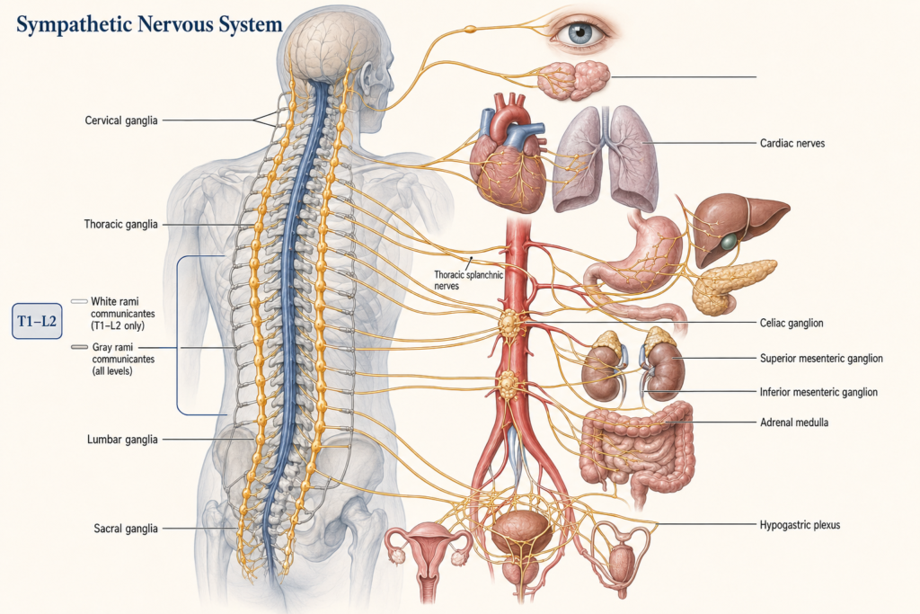
The cell bodies of sympathetic preganglionic neurons sit mainly in a region of the spinal cord called the intermediolateral cell column, from the first thoracic segment, T1, to the second lumbar segment, L2. That is why the sympathetic system is described as having a thoracolumbar outflow.
Most sympathetic signals travel through a two-neuron relay. A preganglionic neuron starts in the spinal cord and sends its axon to a sympathetic ganglion, where it connects with a postganglionic neuron that carries the signal on to the target tissue. The ganglia come in two main groups. Paravertebral ganglia form paired chains running alongside the spine, like two strings of beads. Prevertebral ganglia lie in front of the spine, near the major abdominal arteries. A fiber can connect at the level where it enters the chain, travel up or down the chain before connecting, or pass straight through to a prevertebral ganglion. This flexible wiring lets sympathetic signals reach the head, chest, abdomen, pelvis, and limbs. The bead-chain image only describes the shape, not how signals are routed within it.
The chemical messengers
All sympathetic preganglionic neurons release acetylcholine, which binds mainly to nicotinic receptors on the neurons in the ganglia. Most postganglionic neurons then release norepinephrine, which acts on adrenergic receptors in the target tissue. Different adrenergic receptor types, including alpha and beta receptors, produce different effects depending on the organ.
There are important exceptions, so not every sympathetic nerve uses norepinephrine. The sympathetic fibers supplying most sweat glands release acetylcholine instead. The adrenal medulla works like a modified sympathetic ganglion: preganglionic neurons stimulate its chromaffin cells to release mainly epinephrine, with some norepinephrine, directly into the bloodstream. Nerve signals can therefore produce fast, targeted responses, while hormones from the adrenal medulla produce more widespread effects.
What it does around the body
In the heart, sympathetic stimulation generally increases heart rate, conduction, and the force of contraction, and it also changes the width of selected blood vessels. Together these actions help maintain blood pressure and send blood where it is needed. Sympathetic reflexes are what keep your blood pressure from dropping too far when you stand up.
In the eyes, sympathetic pathways contract the radial muscle of the iris, widening the pupil, a change called mydriasis. They also help lift the upper eyelid slightly through the superior tarsal muscle. In the lungs, sympathetic signaling can relax airway smooth muscle through beta-2 adrenergic receptors, and in humans circulating epinephrine contributes importantly to this effect.
In the skin, sympathetic nerves regulate sweating, skin blood flow, and the tiny muscles that raise body hair, and sweating and skin blood flow are central to temperature control. In the digestive system, sympathetic activity can reduce movement and secretion while tightening some sphincters, temporarily shifting resources away from digestion during physical demand. Digestion isn’t run by one division alone, though. It also depends on parasympathetic pathways, the enteric nervous system, hormones, and local reflexes.
In the bladder, sympathetic pathways help relax the bladder wall and tighten the internal outlet while urine is being stored. Urination itself needs coordinated activity among autonomic, somatic, and central pathways. For metabolism, sympathetic activation helps make energy available by promoting glycogen breakdown, glucose release, and the breakdown of fat in adipose tissue, called lipolysis. The exact response depends on the tissue, receptor type, hormones, and the body’s metabolic state.
Not only for emergencies
The sympathetic system helps the body respond to changing conditions inside and out. In daily life it stabilizes blood pressure when you change posture, adjusts circulation during exercise, supports temperature regulation, controls sweating, regulates energy availability, and coordinates responses to pain, stress, and environmental challenges. Without adequate sympathetic control, simply getting up from lying down could cause a large drop in blood pressure and reduced blood flow to the brain. So the idea that it is active only during danger is wrong.
The fight-or-flight response is real. Facing a perceived threat or major challenge, sympathetic activation can speed and strengthen the heartbeat, widen the pupils, increase sweating, mobilize stored energy, and temporarily reduce digestion. But it is a simplified description. Sympathetic activity also rises during ordinary exercise, exposure to cold, changes in posture, and the period around waking up. And the response is not identical everywhere. Different sympathetic pathways can be regulated separately rather than firing as one uniform system.
How it compares with the parasympathetic system
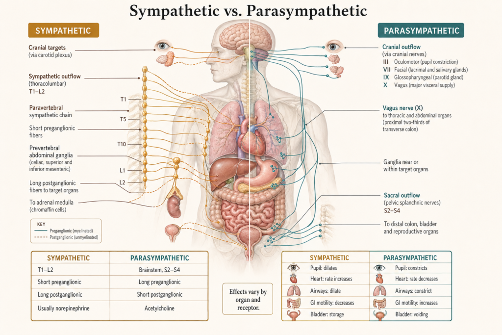
The sympathetic system’s outflow comes from the thoracic and upper lumbar spinal cord, while the parasympathetic system’s comes from the brainstem and sacral spinal cord. Sympathetic ganglia lie near the spinal cord or major abdominal arteries, while parasympathetic ganglia lie near or within the target organs. Both use acetylcholine at the first relay, but the sympathetic system’s main postganglionic messenger is usually norepinephrine, while the parasympathetic system uses acetylcholine. Broadly, the sympathetic side handles mobilization, control of blood vessels, and adaptation to demand, and the parasympathetic side handles maintenance, digestion, and restoration.
That comparison is useful but not a perfect opposition. The two often have opposite effects on heart rate, yet some tissues are controlled mainly by one division, and many responses need cooperation between several systems.
When sympathetic control goes wrong
Problems in sympathetic pathways cause different symptoms depending on where and why they occur. In orthostatic hypotension, sympathetic reflexes fail to tighten blood vessels enough when a person stands, so blood pressure falls, causing lightheadedness, blurred vision, weakness, or fainting.
Damage to the sympathetic pathway serving one side of the face can produce Horner syndrome: a smaller pupil, mild drooping of the upper eyelid, and in some cases reduced sweating on that side. Its cause needs to be investigated, because the damage can occur anywhere between the brain and the eye. Abnormal sympathetic control can also cause excessive, reduced, or uneven sweating, though these symptoms can have neurological, endocrine, medication-related, or other causes.
Sympathetic dysfunction occurs in conditions such as autonomic neuropathies, pure autonomic failure, and some forms of dysautonomia. Symptoms alone can’t show whether sympathetic activity is too high or too low. Clinical evaluation, and in selected cases autonomic testing, is needed. Even a fast heartbeat doesn’t prove the sympathetic system is overactive. It can come from exercise, fever, dehydration, pain, medications, anemia, endocrine conditions, or an abnormal heart rhythm.
This article is educational background, not a diagnostic guide. Fainting, persistent dizziness on standing, or a new drooping eyelid with a small pupil should be evaluated by a clinician.
Related: What Is the Autonomic Nervous System? · What Is the Parasympathetic Nervous System? · What Is the Peripheral Nervous System? · What Is a Neurotransmitter? · How Does the Brain Control the Autonomic Nervous System?
References
- Waxenbaum JA, Reddy V, Das JM. Anatomy, Autonomic Nervous System. In: StatPearls [Internet]. Treasure Island (FL): StatPearls Publishing; 2026. Updated December 1, 2025. NCBI Bookshelf
- McCorry LK. Physiology of the autonomic nervous system. American Journal of Pharmaceutical Education. 2007;71(4):78. PubMed Central
- LeBouef T, Yaker Z, Whited L. Physiology, Autonomic Nervous System. In: StatPearls [Internet]. Treasure Island (FL): StatPearls Publishing; 2026. Updated May 1, 2023. NCBI Bookshelf
- Coon EA. Overview of the Autonomic Nervous System. MSD Manual Professional Edition. Full review May 2025; updated November 2025. MSD Manual
Written by: MedMaru Editorial Team
Reviewed for medical accuracy by:
S. Chang, KMD