What Are Glial Cells? Types and Functions
Neurons get most of the attention, but they cannot run the nervous system by themselves. They depend on a varied group of partner cells called glial cells, also known as glia or neuroglia. Glial cells are the non-neuronal cells that maintain, protect, and regulate the nervous system. They control the chemical environment around neurons, form myelin, support synapses, take part in immune defense, guide development, and respond to injury. They were long regarded as passive support cells, but research now shows they are active participants in how the nervous system works. Normal signaling depends on constant communication between neurons and glia.
More than glue
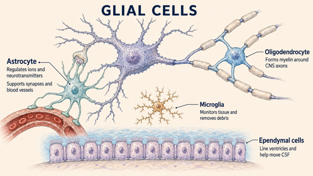
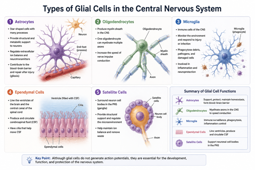
The word glia comes from a word meaning glue, and for a long time that is roughly how these cells were viewed. The name undersells them. Glial cells are found throughout the central nervous system, or CNS, made up of the brain and spinal cord, and the peripheral nervous system, or PNS. The main CNS glia are astrocytes, oligodendrocytes, microglia, and ependymal cells. The PNS has Schwann cells, satellite glial cells, enteric glia in the gut, and other specialized populations.
If neurons are the musicians of an orchestra, glia are the stage crew, sound engineers, and security staff who make the performance possible. The comparison helps, but it undersells glia in one way: they also shape the music, influencing which signals pass and how strongly. Most mature glial cells do not send long-distance action potentials the way neurons do, but they are not electrically or chemically silent. They respond to neurotransmitters, shifts in ion levels, tissue damage, and signals from other cells. Some communicate through calcium signaling and release molecules that affect their neighbors.
You may have heard that glia outnumber neurons ten to one. That claim is outdated. Modern cell-counting studies suggest the adult human brain contains roughly comparable total numbers of neurons and non-neuronal cells. The ratio varies a great deal by region, though. The cerebellum is packed with neurons, while some areas of the cerebral cortex and white matter contain proportionally more glia, so no single ratio describes the whole brain.
Astrocytes: managing the neighborhood
Astrocytes are star-shaped cells found throughout the brain and spinal cord, and their many branches touch neurons, synapses, blood vessels, and other glia. One of their main jobs is keeping the fluid around neurons in balance. Neural signaling needs tightly controlled ion levels, and astrocytes help regulate extracellular potassium and other substances that shift during activity. They also clear neurotransmitters, including glutamate, from around synapses, which limits excessive receptor activation and resets the synapse for the next signal.
Astrocytes also support neuronal energy use. They take up nutrients from the blood, store glycogen, and can supply metabolic fuel when neural activity rises. Exactly how energy passes between astrocytes and neurons is still being researched, so describing astrocytes as cells that simply feed neurons is too simple.
At synapses, astrocytes can influence synapse formation, transmitter clearance, ion balance, synaptic maintenance, and some forms of plasticity. The idea of a tripartite synapse captures this three-way partnership between the sending neuron, the receiving neuron, and the astrocyte’s processes, although astrocyte involvement differs among brain regions and synapse types.
Astrocytes also wrap many CNS blood vessels with their endfeet and help maintain the conditions the blood–brain barrier needs. The barrier’s main physical seal, however, is formed by tight junctions between specialized endothelial cells lining the vessels. Astrocytes support and regulate it rather than create it. After infection, trauma, stroke, or other CNS damage, astrocytes can become reactive, a process called reactive astrogliosis, changing their structure, gene expression, and activity. Reactive astrocytes may help contain injury, restore the local environment, and protect surviving tissue. In some settings, though, glial scarring and related responses can also limit axon regrowth, depending on the type, severity, and timing of the injury.
Oligodendrocytes and Schwann cells: the myelin makers
Myelin is not made by a single cell type. In the brain and spinal cord, oligodendrocytes produce it. Myelin is a lipid-rich membrane wrapped around segments of certain axons. It reduces current loss and lets action potentials travel quickly between gaps called nodes of Ranvier, a pattern known as saltatory conduction. A single oligodendrocyte can extend several arms and myelinate segments of several axons. Oligodendrocytes also give axons metabolic and structural support, so damage to them can disrupt axons as well as myelin.
In peripheral nerves, Schwann cells take over. A myelinating Schwann cell wraps its membrane around a segment of one axon, speeding up conduction. Not every peripheral axon is myelinated, and nonmyelinating Schwann cells surround and support groups of small axons without forming compact myelin around each. After peripheral nerve injury, Schwann cells change their behavior, help clear damaged material, and create an environment that supports regrowth. They don’t guarantee full recovery, though. Regeneration still depends on where and how badly the nerve was injured, the distance to the target, scarring, and whether the axon can reconnect with the right tissue.
Microglia: the resident immune cells
Microglia are the main resident immune cells of the central nervous system, and they have an unusual origin. Astrocytes, oligodendrocytes, and ependymal cells mostly arise from neural lineage cells, but most microglia come from early embryonic myeloid precursors associated with the yolk sac and enter the developing nervous system before birth.
Microglia constantly extend and retract their processes to survey the surrounding tissue, so the old term “resting microglia” is misleading. When conditions change, they can alter their shape, movement, gene expression, and function. Depending on the situation, they may remove damaged cells and debris, detect pathogens or injury, release inflammatory or regulatory molecules, communicate with neurons and other glia, and help with repair. Their responses are diverse and can’t always be sorted neatly into good and bad activation states. Microglia also take part in normal brain development, interacting with developing cells and helping remodel synaptic connections. That doesn’t mean they independently decide which connections or memories survive. Remodeling results from interactions among neural activity, molecular signals, microglia, astrocytes, and other cells.
The other glia
Ependymal cells line the brain’s ventricles and the central canal of the spinal cord. Many have cilia that help move cerebrospinal fluid along these surfaces. Cells related to the ependyma are also associated with the choroid plexus, where cerebrospinal fluid is produced by a structure containing specialized epithelial cells, blood vessels, and connective tissue. It is more accurate to say ependymal cells take part in the ventricular and fluid system than to say ordinary ependymal cells alone produce all cerebrospinal fluid.
Satellite glial cells surround the cell bodies of neurons in sensory and autonomic ganglia outside the brain and spinal cord. They help regulate the local chemical environment and take part in metabolic support and signaling. Changes in these cells are being studied in persistent pain and peripheral nerve disorders, though many mechanisms are still unclear.
Radial glial cells are especially important before birth. They act as progenitors that generate neurons and several kinds of glia, and their long processes serve as scaffolds along which newborn neurons migrate to their destinations. Some specialized radial glia remain in the mature nervous system, including Bergmann glia in the cerebellum and Müller glia in the retina. Oligodendrocyte precursor cells, which can generate new oligodendrocytes and take part in local signaling, and enteric glia, which support neural circuits and tissue balance in the digestive tract, round out the picture.
A two-way conversation
Communication between neurons and glia runs in both directions. Neuronal activity changes the levels of neurotransmitters and ions outside cells. Glia detect these changes and respond by adjusting ion balance, clearing transmitters, changing metabolic support, modifying myelin, or releasing signaling molecules. Glia also talk to each other through chemical messengers, gap junctions, calcium signals, contact-dependent signals, cytokines, and growth factors. How they respond depends on the cell type, brain region, stage of development, and physiological condition, so glia should not be treated as one uniform population.
Taken together, glia maintain ion and transmitter balance, support energy metabolism, form and maintain myelin, regulate synapses, support blood vessels and fluid boundaries, guide development, watch for infection and damage, clear debris, coordinate injury responses, and support peripheral nerve regeneration. A neuron may generate the electrical signal, but it cannot work normally without glial regulation.
Glia in disease
Glial problems contribute to many neurological diseases, though glia are rarely the only cells involved. In multiple sclerosis, immune-mediated inflammation damages CNS myelin and oligodendrocytes, disrupting conduction and possibly leading to axonal injury over time. Schwann cells and peripheral myelin are damaged in conditions such as Guillain–Barré syndrome and some inherited neuropathies.
Gliomas are tumors classified by features resembling particular glial lineages, with major categories including astrocytomas, oligodendrogliomas, and ependymomas. Modern classification also uses molecular and genetic findings, and a tumor’s name doesn’t necessarily mean it arose from a fully mature glial cell of that type.
Astrocytes and microglia also change their behavior in Alzheimer’s disease, Parkinson’s disease, amyotrophic lateral sclerosis, infections, epilepsy, stroke, and traumatic injury. These responses may protect tissue at some stages and harm it at others, so not all glial activation damages the brain. Seeing activated glia in diseased tissue does not by itself prove that glia caused the disease.
This article is educational background and does not replace medical evaluation.
Related: What Is a Neuron? · What Is a Sensory Neuron? · What Is a Motor Neuron? · What Is an Interneuron? · What Is a Synapse? · What Are Neurotransmitters? How Brain Cells Communicate
References
- von Bartheld CS, Bahney J, Herculano-Houzel S. The search for true numbers of neurons and glial cells in the human brain: a review of 150 years of cell counting. Journal of Comparative Neurology. 2016;524(18):3865–3895. doi:10.1002/cne.24040. PubMed
- Ginhoux F, Greter M, Leboeuf M, et al. Fate mapping analysis reveals that adult microglia derive from primitive macrophages. Science. 2010;330(6005):841–845. doi:10.1126/science.1194637. PubMed
- Allen NJ, Lyons DA. Glia as architects of central nervous system formation and function. Science. 2018;362(6411):181–185. doi:10.1126/science.aat0473. PubMed Central
- Fields RD, Stevens-Graham B. New insights into neuron-glia communication. Science. 2002;298(5593):556–562. doi:10.1126/science.298.5593.556. PubMed
Written by: MedMaru Editorial Team
Reviewed for medical accuracy by:
S. Chang, KMD