What Is Myelin? Myelin Sheath Structure and Function
Myelin is a specialized membrane wrapped around many of the nervous system’s axons, the long fibers that carry signals away from nerve cells. It works as electrical insulation, helping nerve signals travel quickly and reliably between the brain, spinal cord, peripheral nerves, muscles, and sense organs. Myelin is rich in lipids and covers an axon in segments, leaving small gaps called nodes of Ranvier where the signal is regenerated. In the brain and spinal cord it is made by cells called oligodendrocytes, and in the peripheral nerves by Schwann cells. When myelin is damaged, nerve signals can slow down or fail, and neurological symptoms can follow.
A living wrapper, not a layer of fat
Myelin is a multilayered extension of the cell membrane of specialized glial cells, the support cells of the nervous system. These membrane layers wrap around part of an axon again and again and become tightly compacted into the myelin sheath. Myelin contains a high proportion of lipids, including cholesterol, phospholipids, and glycolipids, along with proteins that hold its structure together. It is not simply body fat around a nerve. It is a highly organized membrane that living glial cells produce, maintain, and remodel.
Not every axon is myelinated. Some small axons conduct signals without compact myelin, and others are supported by glial cells without being individually wrapped. How much myelin an axon has varies with its type, diameter, location, and function.
Segments and gaps
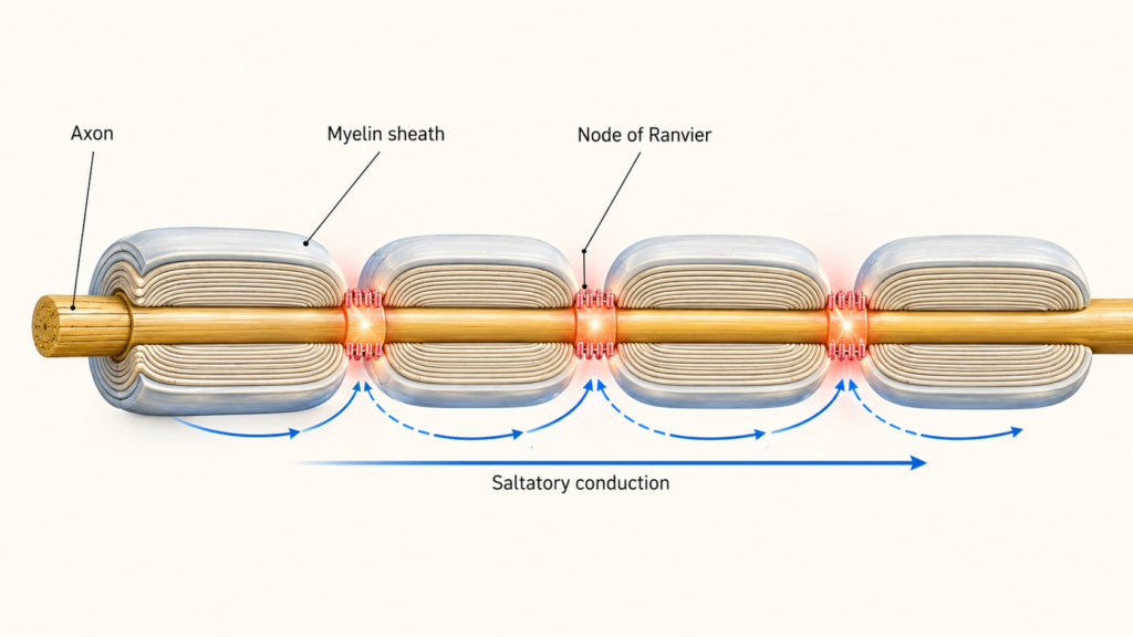
Myelin does not form one continuous tube along the axon. It is laid down in separate sections called internodes, with short gaps between them called nodes of Ranvier. The nodes are packed with voltage-gated sodium channels that regenerate the action potential as it travels. Where the ends of each myelin segment attach closely to the axon, next to the node, is a region called the paranode. Picture a string of sausages along a cord, with the bare cord showing between each link. The picture captures the segmented layout, though real internodes are tightly wound membrane rather than solid tubes.
Two cell types, two styles
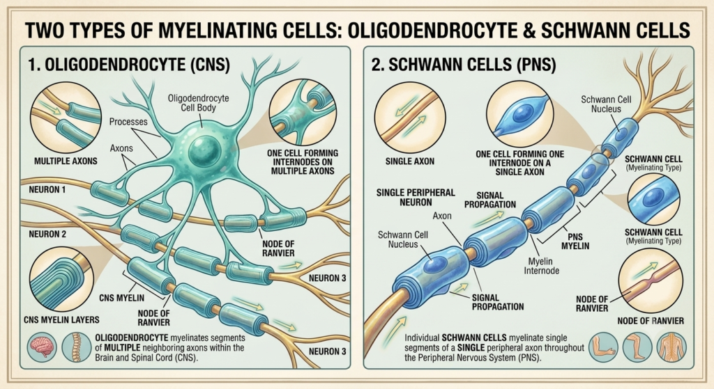
In the brain and spinal cord, oligodendrocytes make myelin. A single oligodendrocyte extends several arms and forms myelin segments on parts of multiple axons. These cells also help support the metabolic and structural health of the axons they touch.
In peripheral nerves, Schwann cells do the job differently. One myelinating Schwann cell normally forms a single segment on one part of one axon, so a long peripheral axon needs many Schwann cells along its length. Some Schwann cells are nonmyelinating. They surround and support groups of small axons without wrapping each one in compact myelin.
Central and peripheral myelin perform similar insulating work, but they differ in cell of origin, molecular makeup, organization, and response to injury. Regeneration of axons after injury is generally limited in the central nervous system and more favorable in peripheral nerves under the right conditions. The two are also linked to different diseases, with multiple sclerosis affecting the central nervous system and some forms of Guillain–Barré syndrome affecting peripheral nerves.
How myelin speeds up signals
An action potential is a rapid electrical change that travels along the axon’s membrane. In many unmyelinated axons it has to be regenerated continuously along each stretch of membrane. Myelin changes the axon’s electrical properties. It increases resistance across the membrane, reduces current leakage, and lowers membrane capacitance, so that local current can spread farther beneath each myelin segment.
When an action potential reaches a node of Ranvier, current travels quickly beneath the next internode and brings the following node to threshold, where a new action potential fires. This is called saltatory conduction, and the signal is often described as jumping from node to node. That description is a simplification. The signal does not leap through empty space. Current spreads beneath the myelin, while action potentials are actively regenerated at each node. Myelin also does not create the nerve impulse itself. It lets action potentials travel faster, more efficiently, and more reliably.
Myelin is a major influence on conduction speed, but not the only one. Speed also depends on axon diameter, myelin thickness, internode length, node structure, the distribution of ion channels, temperature, and axonal health. So not all myelinated axons conduct at the same speed.
Why timing matters
The nervous system cares not only whether a signal arrives, but when. Precise timing lets signals from different neurons and brain regions work together. Myelin supports rapid communication over long distances, reliable sensory processing, coordinated muscle activity, precise timing within circuits, communication between brain regions, efficient organization of pathways, and the metabolic and structural health of axons. It also lets fairly narrow axons conduct quickly. Without myelin, many axons would need to be much wider to reach similar speeds.
In the brain, large bundles of myelinated axons give white matter its lighter color. The corpus callosum and the internal capsule are examples of major white-matter pathways. White matter is not made of myelin alone, though. It also contains axons, glial cells, blood vessels, extracellular material, and some unmyelinated fibers. Gray matter, which holds many cell bodies, dendrites, and synapses, can contain myelinated axons too. Myelin is concentrated in white matter but not limited to it.
Myelin across the lifespan
Myelination starts before birth in some pathways and continues rapidly through infancy and childhood. Different parts of the nervous system become myelinated at different times. Many sensory and motor pathways are myelinated relatively early, while some pathways associated with higher cognitive functions mature later. White-matter development can still be detected through adolescence and early adulthood, although changes seen on brain imaging don’t necessarily reflect myelin formation alone.
Myelin was once seen as fixed insulation, but research now shows that some of it can change in adulthood. Neural activity may influence how oligodendrocytes develop, whether new myelin segments form, which axons get myelinated, the thickness or length of some segments, and the timing of communication within circuits. This is called adaptive myelination or myelin plasticity, and experimental studies suggest it contributes to some forms of motor learning. That does not mean every learning experience creates new myelin, or that memories are stored in myelin. Learning involves coordinated changes in synapses, neurons, glial cells, and circuits.
When myelin is damaged
Loss of or damage to myelin is called demyelination. When myelin is disrupted, current leaks across the axon’s membrane, and signals may slow down, fall out of sync, require more energy, fail to reach the next node, or stop altogether. Symptoms depend on where the damage is and how severe it is, and they can include weakness, altered sensation, vision problems, poor coordination, or cognitive changes. Demyelination does not necessarily mean the axon itself has been destroyed. Persistent inflammation and myelin damage can, however, eventually lead to secondary injury of the axon.
Repair is possible to a degree. The formation of new myelin after damage is called remyelination. In the central nervous system, oligodendrocyte precursor cells can mature into new oligodendrocytes and lay down replacement myelin, which may restore conduction and protect the axon, though repair can become incomplete after repeated or chronic injury. In peripheral nerves, Schwann cells clear damaged tissue, support surviving axons, and form new myelin. Peripheral axons regenerate better than central ones when the cell body survives and the Schwann-cell pathways are sufficiently intact. Recovery still depends on the location, type, and severity of the injury.
Several diseases involve myelin. Multiple sclerosis is an immune-mediated disease involving inflammation and damage in the central nervous system. Myelin is an important target, but MS can also damage axons and other structures, so it is not a disease of myelin alone. Guillain–Barré syndrome is an acute immune-mediated disorder of peripheral nerves. Some forms mainly damage peripheral myelin and structures associated with Schwann cells, while others primarily affect axons, so it is not a single, purely demyelinating condition. Genetic changes affecting myelin proteins, lipid metabolism, or myelin-forming cells can also cause inherited disorders, including certain leukodystrophies, Charcot–Marie–Tooth disease, and Pelizaeus–Merzbacher disease. These differ greatly in cause, symptoms, age of onset, and progression.
This article is educational background, not a diagnostic guide. New weakness, numbness, vision changes, or coordination problems should be evaluated by a clinician.
Related: What Is a Neuron? · What Are Glial Cells? · What Is an Action Potential? · What Is the Central Nervous System? · What Is the Peripheral Nervous System?
References
- Simons M, Nave KA. Oligodendrocytes: myelination and axonal support. Cold Spring Harbor Perspectives in Biology. 2016;8(1):a020479. doi:10.1101/cshperspect.a020479. PubMed Central
- Salzer JL. Schwann cell myelination. Cold Spring Harbor Perspectives in Biology. 2015;7(8):a020529. doi:10.1101/cshperspect.a020529. PubMed Central
- National Institute of Neurological Disorders and Stroke. Multiple Sclerosis. NINDS
- Bellanti R, Rinaldi S. Guillain–Barré syndrome: a comprehensive review. European Journal of Neurology. 2024;31(8):e16365. doi:10.1111/ene.16365. PubMed
Written by: MedMaru Editorial Team
Reviewed for medical accuracy by:
S. Chang, KMD