How Does the Brain Process Vision?
Your Eyes Don’t Send the Brain a Picture
The intuitive model of vision goes something like: eyes work like a camera, capture an image, and send it to the brain to “look at.” That model breaks down almost immediately. The retina doesn’t transmit a picture — it transmits a stream of processed signals, and the image you experience is assembled from those signals well after they leave the eye.
The cornea and lens do focus light onto the retina, the way a camera lens focuses light onto film. But the retinal image is only a starting point, not a finished product. What the eye captures is upside-down and reversed left-to-right — a fact that surprises people less because of any correction happening in the eye, and more because vision was never “a picture” for the brain to flip in the first place. Perception is built from patterns of neural activity and relationships between signals, not from rotating an internal photograph.
Processing starts before the signal even leaves the eye
Inside the retina, rods and cones convert light into electrical activity — rods handling dim light and peripheral vision, cones handling daylight vision, fine detail, and color. That signal doesn’t travel straight to the brain. It passes through a small relay of retinal cells — bipolar, horizontal, and amacrine cells — that shape and organize it before it ever reaches a retinal ganglion cell, whose axon actually carries the signal out of the eye. Meaningful visual processing is already underway before the information leaves the eyeball.
Why damage to one side of the brain affects both eyes
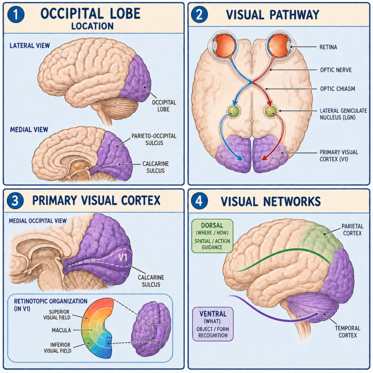
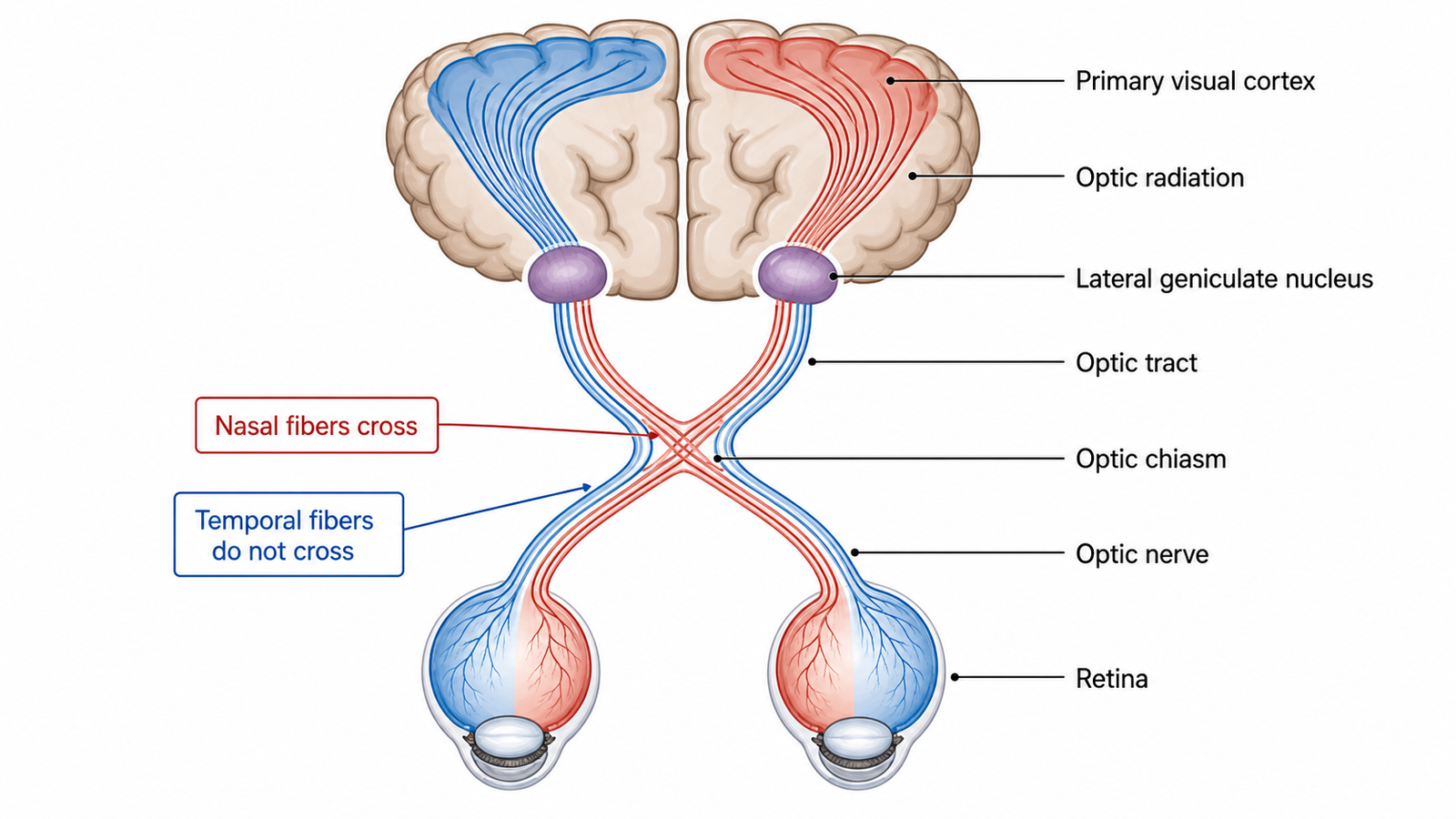
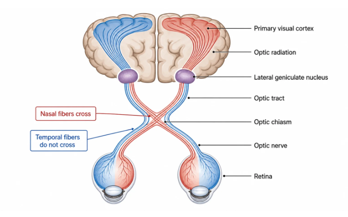
Ganglion cell axons bundle into the optic nerve, and at a junction called the optic chiasm, something unintuitive happens: fibers from the inner (nasal) half of each retina cross to the opposite side of the brain, while fibers from the outer (temporal) half stay on the same side. The result is that each hemisphere doesn’t handle one eye — it handles one half of the visual world, using input from both eyes. The left hemisphere processes what’s happening in your right field of view; the right hemisphere processes the left. This is also why damage to different points along the pathway produces distinct patterns of vision loss — retinal or optic nerve disease can affect just one eye, chiasm damage can disrupt outer visual fields, and damage further back can knock out the same side of vision in both eyes at once. (Sudden vision loss of any kind needs urgent medical evaluation — this kind of pattern recognition is educational, not diagnostic.)
The image gets rebuilt, not just relayed
From the chiasm, most signals reach the lateral geniculate nucleus in the thalamus, which preserves the spatial map of the visual field and routes information onward to primary visual cortex (V1), tucked around the calcarine fissure in the occipital lobe. Even here, neurons aren’t reconstructing a scene — they respond to specific organized features like edges and orientations. It’s only through the surrounding and higher visual areas that these fragments combine into color, motion, depth, faces, and recognizable objects.
That combination work splits, broadly, into two interacting systems: one contributing more to identifying what something is, the other to where it is and how to act on it. These are useful labels for broad, overlapping networks — not two separate wires running in parallel. And the process isn’t strictly one-directional: attention, memory, and expectation feed back into how incoming signals get interpreted, which is part of why perception can differ from the raw sensory input.
What this means practically
Vision, in other words, isn’t located in one place and isn’t a passive recording. It’s distributed across the retina, thalamus, occipital cortex, and a wide network of connected regions, actively shaped by attention and prior experience at every stage. That’s also why “where in the pathway the damage occurred” tells clinicians so much about what kind of visual deficit to expect — the system’s architecture is organized enough that different failure points produce recognizably different symptoms.
Related: How Does the Brain Process Pain? · How Does the Brain Process Sensory Information? · How Does the Brain Maintain Balance?
References
- Gupta M, Ireland AC, Omole AE, Bordoni B. Neuroanatomy, Visual Pathway. StatPearls. Updated March 23, 2026.
- Jozsa F, Hall WA. Neuroanatomy, Retina. StatPearls. Updated February 22, 2026.
- Wurtz RH, McAlonan K, Cavanaugh J, Berman RA. Thalamic pathways for active vision. Trends in Cognitive Sciences. 2011;15(4):177-184.
Written by: MedMaru Editorial Team
Reviewed for medical accuracy by:
S. Chang, KMD