Temporal Lobe: Location, Function, and Anatomy
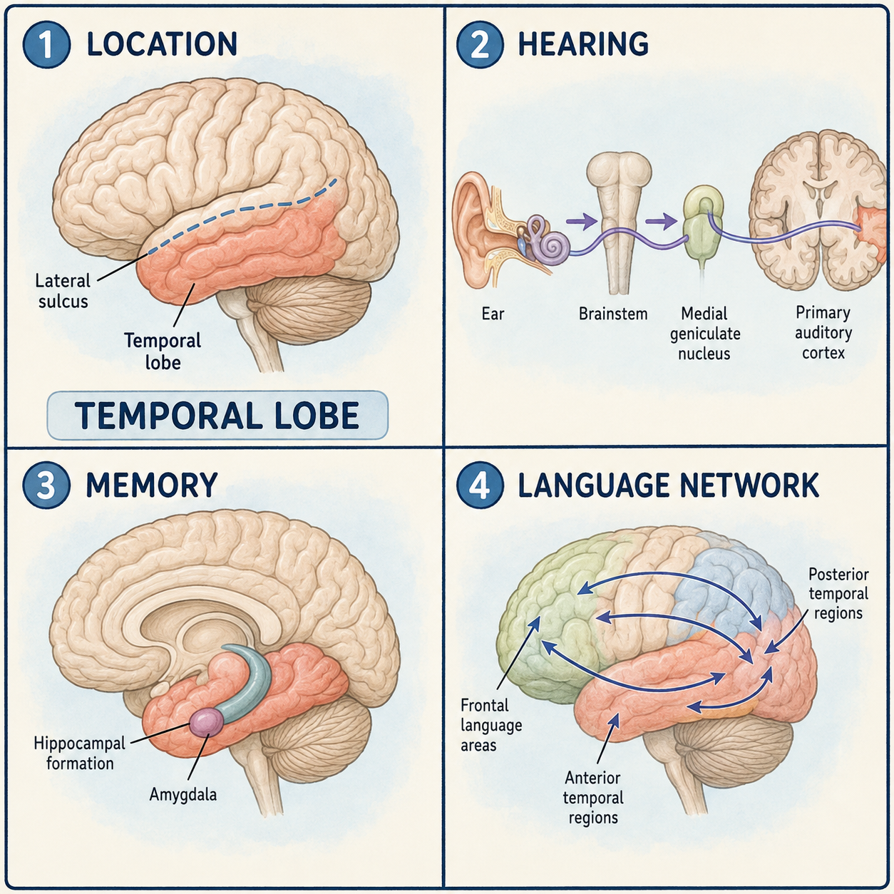
The temporal lobe is the lower side region of each cerebral hemisphere, roughly behind your temple. It lies below a deep groove called the lateral sulcus and contributes to hearing, memory, emotion, object recognition, and language. None of these functions lives in an isolated center. Each one emerges from networks that link temporal cortex with frontal, parietal, limbic, and thalamic structures. So while hearing is a major job here, the temporal lobe is far from only a hearing lobe.
Where it sits and what it is made of
The temporal lobe sits below the lateral sulcus, behind the frontal lobe, and in front of the occipital lobe. Its lower surface rests near the middle cranial fossa, a hollow in the base of the skull. On its outer surface, three long ridges run roughly from front to back: the superior, middle, and inferior temporal gyri. The transverse temporal gyri, also called Heschl’s gyri, hold the primary auditory cortex. They are tucked inside the lateral sulcus, so you cannot fully see them from outside.
Each part of the surface leans toward different work. The superior temporal gyrus includes auditory association cortex, which helps make sense of sound. In the language-dominant hemisphere, its rear portion also takes part in understanding speech. The middle and inferior temporal cortex contribute to semantic knowledge, our store of word and concept meanings, and to recognizing complex objects and visual patterns. Farther underneath, the fusiform gyrus and neighboring ventral temporal cortex take part in high-level visual recognition, including faces and written words, as part of a wider network.
The inner, medial side holds two structures that people often ask about. The hippocampal formation runs along the medial temporal lobe and is essential for forming and organizing new declarative memories, the kind of memories you can consciously recall and describe. The amygdala lies deep in the front, inner part of the lobe and helps assign emotional and motivational significance to what we sense. Both are deep medial temporal structures, so in that sense they are part of the temporal lobe.
The long road from the ear
Sound does not travel straight from the ear to the temporal cortex. The signal passes through several relay stations in the brainstem, climbs through the midbrain, reaches the medial geniculate nucleus of the thalamus, and only then projects to the primary auditory cortex. Along the way, the pathways from each ear spread to both sides of the brain at several levels. Think of it as a message copied to two offices early on, so losing one office doesn’t lose the message. That is why damage to the auditory cortex on one side usually does not cause complete deafness in one ear. The analogy is rough, since the two sides are not perfect duplicates, but it explains the pattern.
Language as a network, not a spot
The classic term Wernicke’s area refers broadly to the rear temporal regions associated with understanding language, usually in the left hemisphere. It is a useful label, but it is not a single fixed spot. Modern imaging and lesion studies show that comprehension, word meaning, reading, and speech production rely on a distributed network that reaches well beyond one patch of cortex, and its exact layout varies between people.
That network has routes running in two directions. Dorsal pathways, including parts of the arcuate fasciculus and the superior longitudinal fasciculus, connect temporal and parietal language regions with frontal cortex. Ventral pathways also contribute, helping map sounds onto meaning. Because of this, conduction aphasia and other language syndromes should not be described as damage to one tract alone. They can reflect injury to a larger network spanning cortex and the white matter beneath it.
A popular shortcut says the left temporal lobe understands language and the right one recognizes faces. That is too simple. Language is left-dominant in most people, but not all. Face recognition uses ventral visual networks on both sides, often with a right-sided emphasis. Memory has a similar shortcut. The hippocampus does not store every memory. It is crucial for forming and organizing many new declarative memories, but long-term memories are represented across distributed cortical networks.
What temporal lobe injury can look like
Damage to the language network in the rear of the dominant temporal lobe can produce fluent speech that is hard to understand, impaired comprehension, or trouble finding words. Damage to medial temporal structures can make it difficult to form new memories, and the exact pattern depends on which side is affected and how widely. Injury to the ventral visual network can impair recognition of objects, faces, or complex visual forms.
Some effects are less obvious. Part of the visual pathway, the optic radiation, loops through the temporal lobe. Damage there causes a loss of the upper quarter of the visual field on the opposite side, sometimes nicknamed a pie-in-the-sky defect. Seizures arising in temporal networks can cause altered awareness, déjà vu, unusual smells, fear, or automatic repetitive movements called automatisms.
These patterns are teaching guides, not diagnostic rules. Symptoms depend on exactly which tissue is involved, which hemisphere is dominant, the vascular territory affected, and the person’s broader brain network.
This article is educational background, not a diagnostic guide. Sudden trouble speaking or understanding speech, or new seizures, needs prompt medical evaluation.
Related: What Is the Cerebral Cortex? · What Is the Brain? · What Is the Cerebrum?
References
Medically reviewed by S. Chang, KMD.
Written by: MedMaru Editorial Team
Reviewed for medical accuracy by:
S. Chang, KMD