
Neuron Structure Explained: Parts of a Nerve Cell and Their Functions
Neuron Structure Explained:
Parts of a Nerve Cell and Their Functions
Introduction
The human brain contains approximately 86 billion neurons, and each one is specialized to receive, process, and transmit information. Every thought, memory, sensation, and movement depends on these remarkable cells communicating with one another.
Although neurons perform many different functions, they all share a similar basic structure. Understanding the anatomy of a neuron makes it much easier to understand how electrical signals travel through the nervous system and how neurological diseases develop.
A neuron consists of several specialized parts that work together to receive and transmit information. The major structures are dendrites, the cell body (soma), the axon hillock, the axon, the myelin sheath, the nodes of Ranvier, and the axon terminals. Each part has a specific role in neural communication.
The Main Parts of a Neuron
A typical neuron can be divided into seven major structures.
- Dendrites
- Cell body (Soma)
- Axon hillock
- Axon
- Myelin sheath
- Nodes of Ranvier
- Axon terminals
Together, these structures allow information to travel rapidly and efficiently throughout the nervous system.
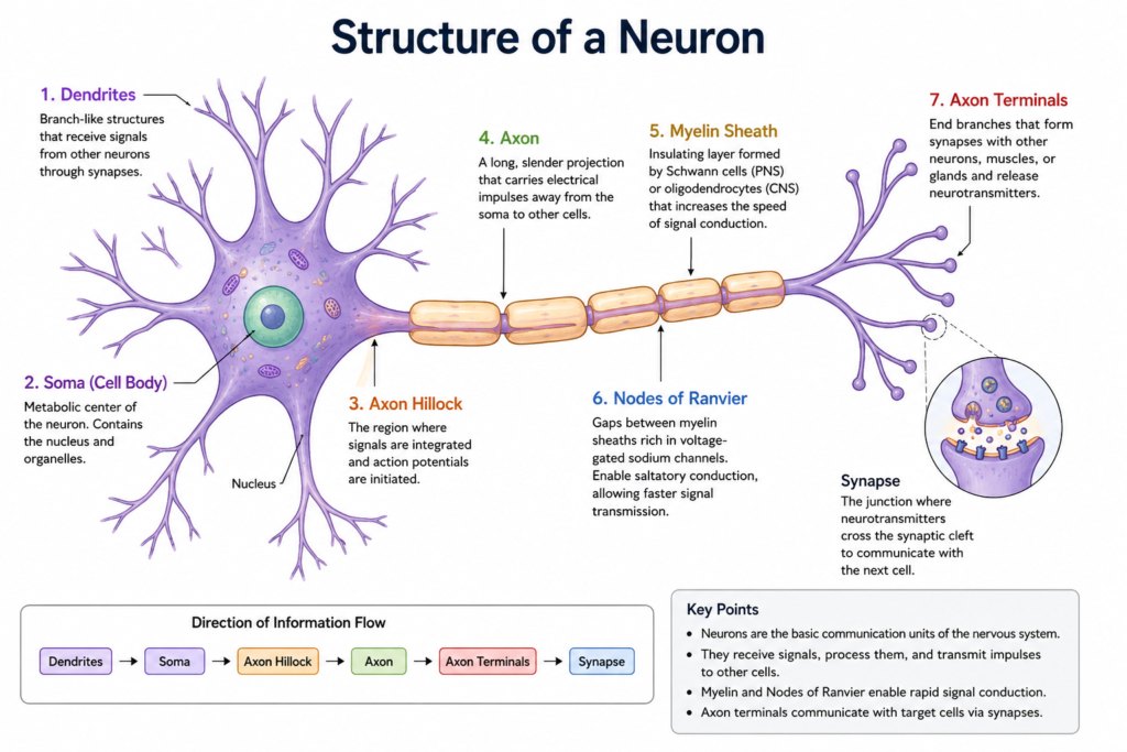
Dendrites
Dendrites are tree-like branches extending from the cell body.
Their primary role is to receive information from other neurons through specialized connections called synapses.
A single neuron may have hundreds or even thousands of dendritic branches, allowing it to receive signals from many different neurons at the same time.
Instead of generating electrical impulses, dendrites collect incoming information and deliver it to the cell body.
Cell Body (Soma)
The cell body, also called the soma, is the metabolic center of the neuron.
It contains the nucleus along with organelles such as mitochondria, ribosomes, the Golgi apparatus, and endoplasmic reticulum. These structures produce proteins, generate energy, and maintain the health of the neuron.
The soma also integrates incoming signals from many dendrites before determining whether an electrical impulse should be generated.
Axon Hillock
The axon hillock is the region where the cell body connects to the axon.
This area acts as the neuron’s decision-making center.
Signals arriving from many dendrites are summed here. If the combined electrical activity reaches a critical threshold, an action potential is generated.
Because it contains a very high density of voltage-gated sodium channels, the axon hillock is usually where nerve impulses begin.
Axon
The axon is a long, thin extension that carries electrical signals away from the cell body.
Some axons are only a fraction of a millimeter long, while others can exceed one meter in length, such as those extending from the spinal cord to the foot.
Unlike dendrites, which mainly receive information, axons transmit information in one direction—from the cell body toward the axon terminals.
Myelin Sheath
Many axons are wrapped in a protective insulating layer called the myelin sheath.
Myelin is rich in lipids and is produced by different cells depending on the location of the neuron.
- In the central nervous system, myelin is produced by oligodendrocytes.
- In the peripheral nervous system, myelin is produced by Schwann cells.
The myelin sheath greatly increases the speed of nerve impulse conduction while reducing the energy required for signal transmission.
Diseases that damage myelin, such as multiple sclerosis, can significantly slow or block nerve conduction.
Nodes of Ranvier
The myelin sheath does not completely cover the axon.
Small gaps called the nodes of Ranvier occur between adjacent segments of myelin.
These nodes contain a very high concentration of voltage-gated sodium channels.
Instead of moving continuously along the membrane, action potentials appear to jump from one node to the next in a process known as saltatory conduction. This allows signals to travel much faster than they would in unmyelinated axons.
Axon Terminals
At the end of each axon are many small branches called axon terminals.
When an action potential reaches these terminals, it triggers the release of neurotransmitters into the synapse.
These chemical messengers then transmit information to another neuron, a muscle cell, or a gland, allowing communication throughout the nervous system.
How Information Flows Through a Neuron
Information travels through a neuron in a specific direction.
Dendrites → Cell body (Soma) → Axon hillock → Axon → Axon terminals → Synapse → Next neuron
Each structure performs a specialized role, making neural communication both rapid and highly organized.
Why Neuron Structure Matters
Every part of the neuron has a unique function.
- Dendrites receive incoming signals.
- The soma integrates those signals.
- The axon hillock initiates action potentials.
- The axon carries electrical impulses.
- The myelin sheath increases conduction speed.
- The nodes of Ranvier regenerate the electrical signal.
- Axon terminals communicate with the next cell using neurotransmitters.
Damage to any of these structures can impair nervous system function and contribute to neurological disease.
Clinical Relevance
Many neurological disorders affect specific parts of the neuron.
| Structure | Examples of disorders |
|---|---|
| Dendrites | Alzheimer’s disease (loss of dendritic spines) |
| Axon | Traumatic axonal injury, peripheral neuropathy |
| Myelin sheath | Multiple sclerosis, Guillain–Barré syndrome |
| Axon terminals | Botulism, Lambert–Eaton myasthenic syndrome |
Understanding neuron anatomy helps explain why these diseases produce characteristic neurological symptoms.
- Neurons are specialized cells that receive, process, and transmit information throughout the nervous system.
- Every neuron has the same basic structural plan, including dendrites, a cell body, an axon, myelin, and axon terminals.
- Each part of the neuron performs a specific function that allows information to flow efficiently in one direction.
- Myelin and the nodes of Ranvier enable rapid signal transmission through saltatory conduction.
- Damage to different parts of a neuron can lead to different neurological disorders.
Related Articles
- What Is a Neuron?
- What Is a Synapse?
- What Is an Action Potential?
- What Are Neurotransmitters?
- How Do Neurons Communicate?
References
- National Institutes of Health. Information about the Brain. NIH Curriculum Supplement Series. NCBI Bookshelf.
- Standring S, ed. Gray’s Anatomy: The Anatomical Basis of Clinical Practice. 42nd ed. Elsevier; 2020.
- Caire MJ, Reddy V, Varacallo MA. Physiology, Synapse. StatPearls. Updated March 27, 2023.
Written by: MedMaru Editorial Team
Reviewed for medical accuracy by:
S. Chang, KMD