Reticular Formation: Location, Function, and Anatomy
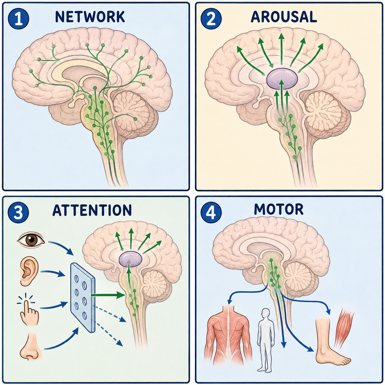
The reticular formation is a loosely woven network of nerve-cell clusters running the full length of the brainstem. It isn’t a neat, boxed-in structure like a lobe or a gland. It looks more like a net stretched from the top of the brainstem to the bottom, which is where the name comes from: the Latin reticulum means “little net.” This net does two very different jobs at once. An upward-reaching part helps keep you awake and alert, while other parts quietly run background tasks such as breathing, heart rate, and muscle tone, mostly without you ever noticing.
A net, not a nucleus
The reticular formation is often imagined as a single nucleus, but it is a diffuse column of more than 100 small clusters of neurons, or nuclei. It runs from the midbrain at the top, through the pons in the middle, down to the medulla oblongata at the bottom, and spills a short way into the upper spinal cord.
Anatomists describe it as three columns arranged from the midline outward. The raphe nuclei sit right on the midline and release serotonin, a messenger linked to mood and the sleep–wake cycle. The medial, or gigantocellular, column has larger cells and coordinates movement and long motor commands. The lateral, or parvocellular, column has smaller cells toward the outer edge and handles local reflexes, including the rhythm of breathing.
A few nuclei within the network have their own names. The locus coeruleus in the pons releases norepinephrine and drives the stress and arousal response. The pedunculopontine nucleus is an acetylcholine-using hub for arousal and REM sleep.
Picture the brainstem as a three-story building, with the midbrain on top, the pons in the middle, and the medulla at the bottom, and the reticular formation as wiring running up the central shaft from floor to floor, with no walls between it and its neighbors. A real net, though, has the same openings everywhere, and this one doesn’t. Its neurons are specialized by region, with the midbrain end leaning toward arousal and the medullary end leaning toward vital reflexes like breathing.
Where it runs, and why that matters
Because the reticular formation has no sharp boundary with the gray matter around it, it shares space with major pathways. The corticospinal tract, carrying movement commands downward, and the spinothalamic tract, carrying pain and temperature signals upward, both run alongside it in the pons and midbrain. A single small brainstem stroke can therefore damage a nearby tract and part of the reticular formation at the same time.
The network connects in both directions. Upward, a set of pathways climbs to the thalamus and on to the cerebral cortex. Downward, the reticulospinal tracts run into the spinal cord, adjusting muscle tone and posture.
How it helps keep you awake
The upward-climbing part has its own name: the ascending reticular activating system, or ARAS. It is the part most directly responsible for wakefulness. It is easy to treat the ARAS and the reticular formation as the same thing, but the ARAS is only the ascending, arousal-related portion of a larger network that also has descending motor and autonomic roles.
Sensory input from the skin, ears, eyes, and internal organs travels through the brainstem toward higher centers, with branches feeding into the reticular formation along the way. Wakefulness then depends on several chemically distinct groups of neurons sending fibers up toward the thalamus and cortex. Norepinephrine comes from the locus coeruleus, serotonin from the raphe nuclei, and acetylcholine from the pedunculopontine nucleus. Two groups in the hypothalamus work alongside them: histamine from the tuberomammillary nucleus and orexin from the lateral hypothalamus. Together, this activating input keeps the thalamus passing sensory information through to the cortex and keeps the cortex’s electrical activity in the desynchronized pattern of wakefulness, rather than the slow, synchronized rhythms of deep sleep. Meanwhile, downward control runs in parallel: reticulospinal tracts adjust muscle tone, and neurons in the medulla help pace breathing and blood pressure.
Think of these systems as separate circuit breakers that help keep the lights of the cortex on. Unlike real breakers, they interact in graded, overlapping ways, and no single chemical acts as the one wakefulness switch. Even orexin, whose loss is linked to narcolepsy type 1, works as one system among several.
When it is damaged
Because the reticular formation, through the ARAS, is one of the main systems keeping the cortex awake, damage to it from a brainstem stroke, severe traumatic brain injury, or certain toxins can cause anything from unusual sleepiness to coma, depending on how much of the network is affected. That is why brainstem injury is treated as a high priority in intensive care: a lesion here can impair consciousness even when the cortex itself is intact. Because circuits for breathing and blood-vessel control sit in the medullary part of the network, injury there can also disrupt breathing rhythm and blood pressure.
It is worth being clear about the limits of current knowledge. Because the reticular formation is diffuse and its many nuclei are deeply interconnected, researchers still have open questions about exactly how each part contributes to arousal versus autonomic control. It is not a fully mapped, settled circuit.
This article is educational background, not a diagnostic guide. Sudden unexplained drowsiness, confusion, or reduced responsiveness needs emergency medical care.
Related: What Is the Cerebral Cortex? · What Is the Brain? · What Is the Brainstem?
References
- StatPearls. “Neuroanatomy, Reticular Formation.” NCBI Bookshelf, NBK556102 — https://www.ncbi.nlm.nih.gov/books/NBK556102/
- StatPearls. “Neuroanatomy, Reticular Activating System.” NCBI Bookshelf, NBK549835 — https://www.ncbi.nlm.nih.gov/books/NBK549835/
Written by: MedMaru Editorial Team
Reviewed for medical accuracy by:
S. Chang, KMD